Home › Skull Base & Craniofacial Tumour Surgery
Craniofacial Surgery in Ahmedabad
Adult craniofacial & skull base tumour surgery · Led by Dr Smit A. Desai, MDS (OMFS)
For tumours of the upper jaw, sinuses, orbit and skull base, we remove the disease completely and rebuild the face in the same operation — protecting your vision, appearance, speech and swallowing. Send your scans and we will tell you honestly whether it can be operated.
Medically reviewed by Dr Smit A. Desai, MDS (OMFS) · Fellowships: Oral Oncology (AOMSI), Head & Neck Oncology (FHNO)
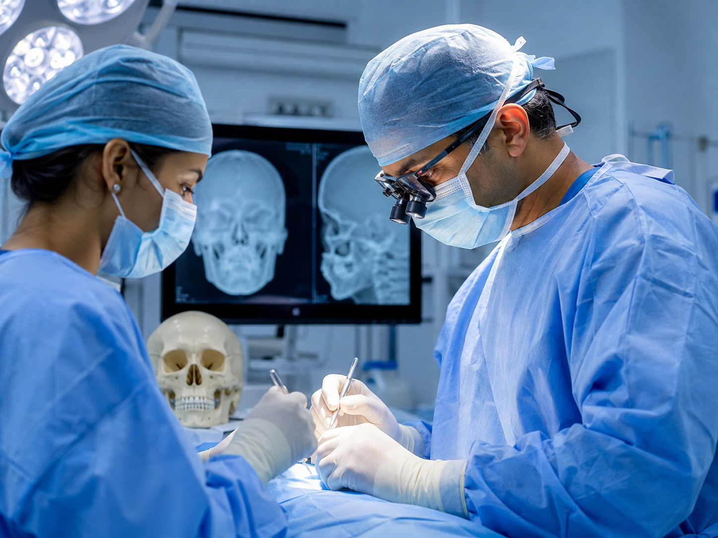
What is craniofacial surgery?
Craniofacial surgery is the surgical treatment of disease affecting the bones and soft tissues of the face, orbit, upper jaw and skull base. In adult cancer care it means removing tumours that involve the sinuses, orbit, palate or skull base, and reconstructing the face in the same operation.
The face is a stack of compartments — sinuses, eye socket, jaws, the deep spaces behind the cheek, and the floor of the skull above them. A tumour in one can push into the next.
Two problems are solved at once: remove the tumour in one piece with clear margins, and protect the sight, bite, speech and face you live with afterwards.
What craniofacial surgery protects — your vision, face, speech and swallowing
Almost nobody facing this operation says “margin”. They ask: will I still see, will I look like myself, will I talk and eat normally?
- Your vision. If the periorbita — the tough lining around the eyeball — is intact, the eye and the sight in it can very often be kept.
- Your face. Craniofacial reconstruction surgery restores cheek projection, eye level and nasal outline, so the face in the mirror is recognisably yours.
- Your speech. The palate is a speech organ. An obturator or a flap closes the gap so speech keeps its resonance instead of turning nasal.
- Your swallowing. Separating mouth from nose again is what lets you drink without fluid coming down your nose.
- The seal between face and brain. At the skull base a watertight barrier is rebuilt so cerebrospinal fluid cannot leak and infection cannot travel upward.
Conditions we treat
If your report names one of these, send it to us and we will explain it in plain language.
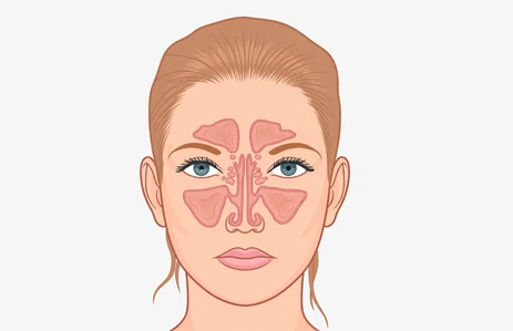
Maxillary sinus & sinonasal cancer
Often mistaken early for ordinary sinusitis.
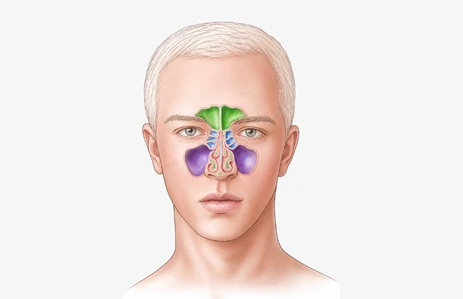
Squamous cell carcinoma of the sinonasal tract
The commonest malignant histology here.
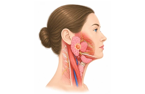
Adenoid cystic carcinoma
A salivary-type cancer that creeps along nerves.
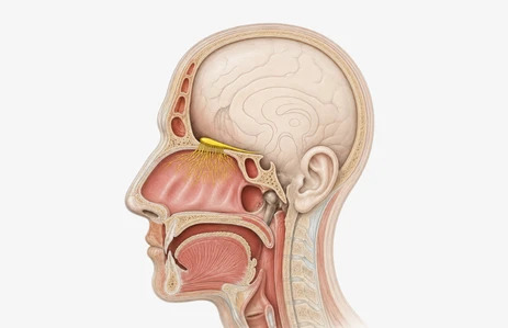
Esthesioneuroblastoma
Olfactory neuroblastoma at the roof of the nose.
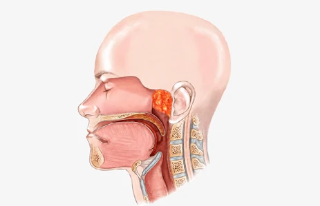
Juvenile nasopharyngeal angiofibroma
Benign, but highly vascular.
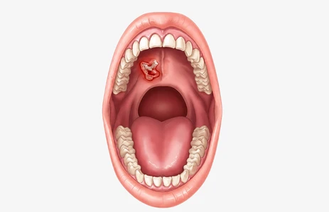
Tumours of the upper jaw & hard palate
Partial or total maxillectomy.
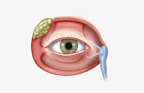
Orbital involvement by tumour
The decision: can the eye be preserved?
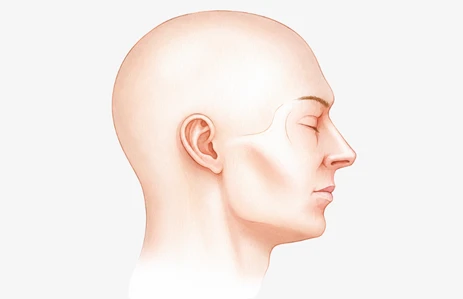
Infratemporal fossa tumours
The deep space behind the upper jaw.
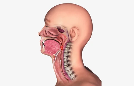
Oral cancer spreading to the skull base
Advanced mouth cancer or tongue cancer travelling backwards, or along nerves.
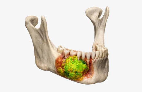
Ameloblastoma & aggressive benign jaw lesions
Benign but locally aggressive; they recur.
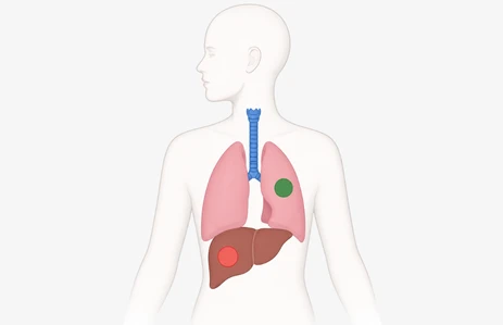
Recurrent tumours in operated or irradiated fields
Salvage surgery — accepted for review.
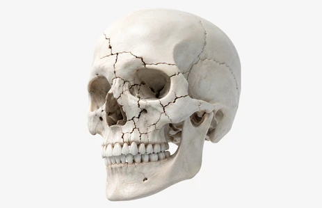
Complex craniofacial trauma
Midface, orbital and panfacial fractures.
Not sure which one your report names? Send the biopsy report and the scan.
ENT skull base surgery — the approaches, and who is in the room
“ENT skull base surgery” is not one operation. It is three routes into the base of the skull, chosen by where the tumour sits.
Endoscopic endonasal approach
Through the nostrils with an endoscope — no facial incision. These endoscopic skull base approaches suit selected tumours of the nasal cavity, ethmoid and central skull base.
Open craniofacial resection
The face is opened from below — the part Dr Smit leads. Where the tumour reaches the dura, a neurosurgeon opens from above in the same operation.
Lateral & transfacial approaches
Through the side of the face, for disease behind the upper jaw or in the masticator space.
Who is actually in the room
ENT skull base surgery is never a one-surgeon operation. Dr Smit leads the maxillofacial, transfacial and reconstructive part. He does not perform craniotomy or intracranial resection.
Head & neck / maxillofacial surgeon
Dr Smit A. Desai — resection, neck and reconstruction plan.
ENT / rhinology skull base surgeon
Leads the endonasal corridor.
Neurosurgeon
Dura and any intracranial component.
Microvascular reconstruction
Free-flap transfer under the operating microscope.
Maxillofacial prosthodontist
Makes the obturator that rebuilds the palate.
Oculoplastic surgeon
Eye preservation, eyelid and socket.
Radiation & medical oncology
Adjuvant treatment, decided before surgery — see diagnosis and treatment of oral and neck cancers.
Pathologist
Frozen sections during the operation, so margins can be extended.
Anaesthesia & critical care
Neuro-aware anaesthesia and intensive-care support.
Craniofacial reconstruction surgery — rebuilding the face after tumour removal
Removing the tumour is the half that gets discussed. Craniofacial reconstruction surgery is the half that decides what life looks like afterwards.
- Microvascular free-flap reconstruction. Living tissue — skin, muscle, and bone where the jaw or orbital rim must be rebuilt — is moved from elsewhere in the body, its artery and vein stitched to neck vessels under an operating microscope. It holds the shape of the face and heals through radiotherapy.
- Orbital and midface reconstruction. Rebuilding the orbital floor and rim keeps the eyeball at the correct level so the eyes still line up; cheek projection stops the face looking collapsed.
- Obturator and prosthetic rehabilitation after maxillectomy. Not every palatal defect needs a flap. A well-made obturator plugs the gap between mouth and nose, restores speech and swallowing quickly, and keeps the cavity visible for follow-up.
- Dental and functional rehabilitation. Chewing is restored through the prosthesis or, in selected cases, implants in reconstructed bone once healing and radiotherapy are complete.
- Soft-tissue detail. Eyelid position, lip competence and cheek contour are planned in the same sitting, not left for a later "revision".
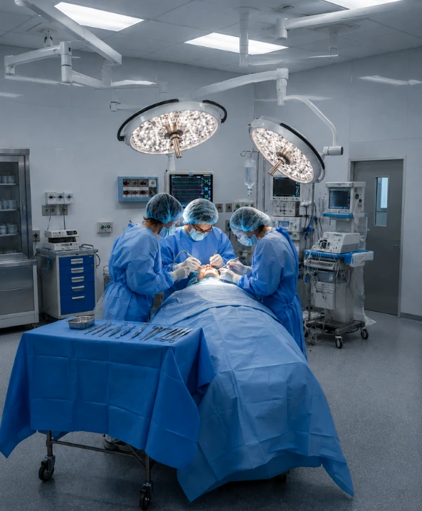
Can the eye be saved? The honest answer
If the tumour has reached the orbital floor but its lining is intact, the eye can usually be preserved. If it has grown through, removal may be the only way to clear the cancer.
Imaging indicates; frozen sections in theatre decide. You hear both possibilities in plain language before you sign anything — including what an orbital prosthesis looks like.
Is the tumour operable? Get a second opinion on your scans
Families arrive carrying a scan and a sentence: “nothing can be done”, or “the eye will have to be removed”. Sometimes that is correct. Sometimes it reflects what one hospital is set up to do.
Dr Smit A. Desai’s published work on compartment resection in advanced buccal mucosa cancer covers exactly the deep spaces that border the skull base — the same ground as surgery for advanced oral and neck cancer.
Send the imaging
WhatsApp the CT, MRI or PET-CT to +91 99983 19470. Photographs of the films are usually enough to begin.
Send the biopsy report
Without histopathology, any opinion on operability is guesswork.
Personal review by Dr Smit
Discussed with ENT skull base surgery and neurosurgical colleagues where disease approaches the skull base. Urgent cases are seen on priority.
An honest answer, in writing
Whether it is operable, what the operation involves, the reconstruction plan, and the real trade-offs — including for the eye.
Sometimes "inoperable" is the truthful answer
Some tumours genuinely cannot be removed with acceptable risk — carotid encasement, extensive brain invasion, or spread to other organs. Saying so is part of the job.
You are not sent away with nothing. There is still a plan: non-surgical oncology, procedures to relieve pain or bleeding, and honest support for the family.
Craniofacial surgery for tumours — who this service is for
This service is for you if:
- You are an adult with a biopsy-proven tumour of the sinus, upper jaw, palate or orbit
- An oral cancer has grown backwards or upwards towards the skull base
- You have been told the tumour is close to the eye, or that the eye may have to be removed
- You have a recurrent tumour in a previously operated or irradiated area
- You need the face rebuilt after a maxillectomy or a large facial resection
- You have complex midface, orbital or panfacial fractures
- A child has a diagnosed cancer of the face, jaws or sinuses — children with cancer are treated here
This service is not for:
- Craniosynostosis or syndromic skull shape in children
- Cleft lip and cleft palate repair, at any age
- Cosmetic facial reshaping without disease
For these conditions we refer families to a dedicated paediatric craniofacial unit.
What affects the cost of craniofacial surgery
There is no single craniofacial surgery cost. Any hospital quoting one before seeing your scans is quoting a number that will change.
The variables that decide your estimate
- Tumour size & skull base involvement — sets the scale of everything.
- Single-team vs two-team surgery — neurosurgery and ENT skull base surgery colleagues.
- Type of reconstruction — closure, local flap, free flap or obturator.
- Theatre & anaesthesia time
- ICU and ward days — one of the largest variables.
- Imaging — CT, MRI, PET-CT.
- Frozen section & histopathology
- Implants, plates & prosthesis
- Whether radiotherapy follows
- Rehabilitation — speech, swallowing, dental.
Get a free written, itemised estimate
Once we have seen your scans and biopsy report, you receive an itemised estimate — surgery, reconstruction, stay and investigations, line by line.
- Itemised in writing, not a verbal figure
- Explained before you decide anything
- Insurance pre-authorisation assistance Cashless and reimbursement claims supported
- No obligation to proceed with us
Why we don't publish a fixed price
Two patients with the same diagnosis on paper can need operations differing by hours, an extra surgical team and a different reconstruction. One published craniofacial surgery cost figure would mislead one of them.
You get an itemised craniofacial surgery cost estimate written for your case once your scans are read — free.
Craniofacial surgery in India — why patients travel to Ahmedabad
Patients ask about craniofacial surgery in India for three honest reasons: surgeons here see advanced disease routinely, the same surgery and reconstruction cost far less than in the West or the Gulf, and the whole pathway sits in one city. Heyansh runs three clinics across Ahmedabad.
Considering craniofacial surgery in India from another state or abroad? Send your scans first.
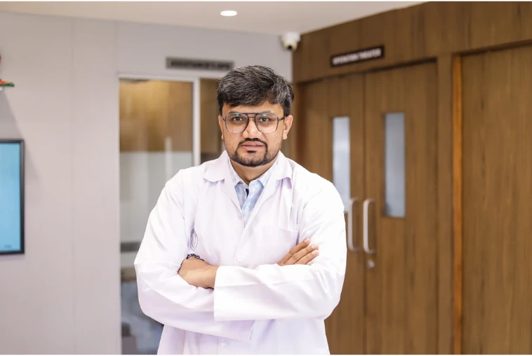
Meet your surgeon — Dr Smit A. Desai
Consultant Oral & Maxillofacial Oncosurgeon · Heyansh Oral Cancer Hospital, Ahmedabad
He also practises as a head and neck cancer surgeon in Ahmedabad. His credibility here is published, not asserted — author of work on advanced buccal mucosa cancer involving the masticator space (T4b) and on outcomes in oral cancers labelled technically unresectable.
Those compartments are the corridor through which oral cancer reaches the skull base. He uses an operating microscope for reconstructive and oncoplastic surgery.
- Junior Consultant, Head & Neck Oncology, Shankus Medicity, Mehsana
- Member — AOMSI · IAOO · IAOMS · FHNO
- Best poster, 6th World Congress 2017, IAOO Bangalore
- Speaker at 20+ national and international conferences
Why choose Heyansh Oral Cancer Hospital for craniofacial surgery
Every line below is a fact you can check.
Published in this anatomy
Peer-reviewed work on compartment resection for advanced buccal mucosa cancer involving the masticator space.
Two oncology fellowships
Oral Oncology (AOMSI first batch, 2017–2019) and Head & Neck Oncology (FHNO, 2018) — cancer training, not general surgical training.
Operating microscope on site
Microvascular free-flap reconstruction, so removal and rebuilding happen in one sitting.
4.9 from 261 Google reviews
Over 10+ years, 8,000+ patients treated and 50–70 surgeries every month.
A real multidisciplinary team
Neurosurgery, ENT skull base surgery, oculoplastics, prosthodontics, pathology and oncology — each role stated openly.
Honest scope
No cleft or craniosynostosis surgery, and no craniotomy by our own surgeon — so you are never referred onward late.
Recurrent & "inoperable" cases reviewed
Salvage surgery in irradiated fields is accepted for assessment. Read a seven-year survivor’s story and an oral cancer recovery at 80.
One pathway, one city
Diagnosis, surgery, reconstruction and follow-up at a dedicated oral cancer hospital in Ahmedabad.
3 clinics across Ahmedabad
Maninagar, Satellite and Nikol/New Naroda — get directions and timings.
Book your consultation
Send your CT/MRI and biopsy report on WhatsApp for an honest opinion on whether the tumour is operable.
Also 087349 54487 (Maninagar) · 098983 48601 (Nikol) · drsmitdesai111@gmail.com · Mon–Sat 10am–2pm & 5–8pm
Craniofacial surgery — frequently asked questions
No. Most craniofacial surgery here is adult tumour work — cancers of the sinuses, upper jaw, orbit and skull base, plus complex facial trauma. Children are treated when the diagnosis is cancer. Cleft lip and palate and craniosynostosis are not performed here.
Not usually. The skull base is the floor the brain rests on, and most tumours treated here sit below it — in the sinuses, nose, orbit or upper jaw. Where disease reaches the brain covering, a neurosurgeon joins the operation.
Often, yes. If the tumour has reached the bone of the orbit but its lining is intact, the eye can usually be preserved. If it has grown through into the orbital fat or the eye itself, removal may be the only way to clear the cancer.
It depends on the route. Selected tumours come out entirely through the nostrils, leaving no facial scar. Larger ones need a facial incision, placed in natural creases wherever possible.
Not necessarily. Sometimes it is accurate; sometimes it reflects what one hospital is set up to do. Send your scans and biopsy report for an independent opinion before accepting it.
The gap between mouth and nose is closed either with an obturator or with a microvascular free flap using your own tissue. Which suits you depends on the defect. Speech and eating are planned before surgery, not after.
A removable prosthesis, made by a maxillofacial prosthodontist, that plugs the defect in the roof of the mouth so you can speak and eat normally.
Tumour size and skull base involvement, whether a second surgical team is needed, the reconstruction type, theatre and ICU time, and whether radiotherapy follows. You get a written itemised estimate once your scans are read — free.
Frequently, yes. For most malignant tumours of the sinuses, upper jaw and skull base, surgery is followed by radiotherapy, sometimes with chemotherapy. The tumour board decides after the pathology report.
A team. Dr Smit A. Desai leads the maxillofacial, transfacial and reconstructive part; an ENT colleague leads endoscopic endonasal work and a neurosurgeon handles anything intradural. He does not perform craniotomy or intracranial resection.
Craniofacial surgery in Ahmedabad — three clinics across the city
Heyansh Oral Cancer Hospital · Dr Smit A. Desai · trusted by families across Gujarat
Maninagar
305, Shreekar Avenue, Old Chawla Park Society, nr Bhairavnath BRTS, Gopal Chowk, Maninagar, Ahmedabad 380008
Satellite
Santorini Square, Abhishree Complex, 512, Satellite, Ahmedabad 380015
Mon–Sat 10am–2pm & 5–8pm · Sun closed
Nikol / New Naroda
Rosevill Sky A, A/33, Nikol–Naroda Rd, opp. Pushkar Icon, New Naroda, Nikol, Ahmedabad 382345
Mon–Sat 10am–2pm & 5–8pm · Sun closed
This page is general information and does not replace a medical consultation — learn more from the National Cancer Institute.