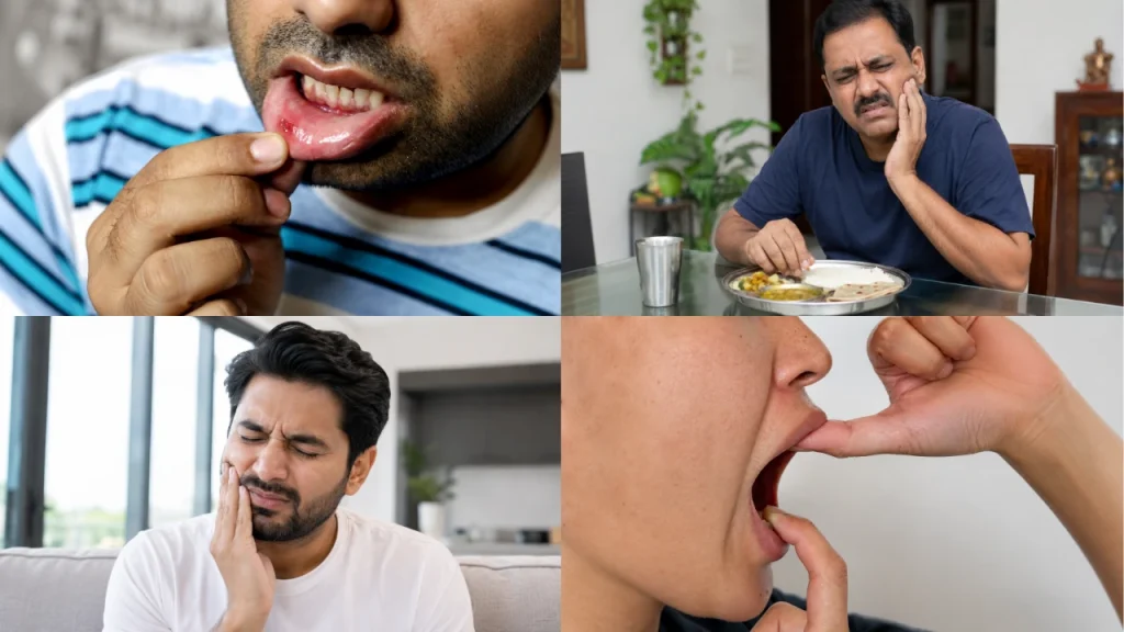
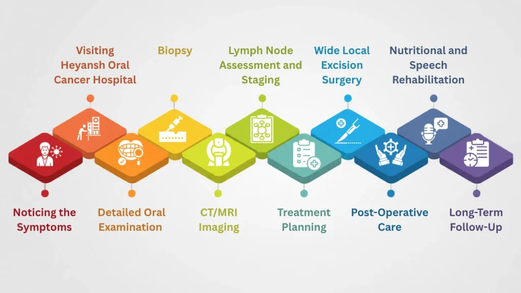
Author: Dr Smit

After surgery

Understanding Your Oral Cancer Pathology Report: Margins, Nodes and Grade

Your oral cancer pathology report is the document that decides whether surgery was enough, or whether more treatment is advised. Here is what each line on it means, in plain English.
Written by the clinical team at Heyansh Oral Cancer Hospital, Ahmedabad
Medically reviewed by Dr Smit A. Desai, MDS, Fellow in Oral Oncology
Published 28 October 2026 · Last reviewed 28 October 2026 · 12 min read

Key takeaways

  • The oral cancer pathology report after surgery is a different, fuller document from the biopsy report that confirmed your diagnosis.
  • Margins describe the rim of normal tissue around the tumour: over 5 mm is usually called clear, 1–5 mm close, and under 1 mm involved.
  • Lymph nodes are reported as a fraction, such as 1 out of 28. Cancer breaking through a node’s outer capsule is called extranodal extension, and it matters a great deal.
  • Grade describes how closely the cancer cells still resemble normal ones. It is one factor among several, not a verdict on its own.
  • No single line decides anything. The findings are read together to decide whether radiotherapy, or radiotherapy with chemotherapy, is advised after surgery.

What is an oral cancer pathology report?

An oral cancer pathology report is the written result of a pathologist examining everything the surgeon removed — the tumour, the rim of normal tissue around it, and any neck lymph nodes — under a microscope. It confirms the type of cancer, measures how far it had spread, and states whether it was removed completely. It is the document that decides whether surgery alone was enough.

In India the document is usually headed histopathology report, and your surgeon may call it “the final HPR”. It is written for another doctor, not for you, which is why it reads the way it does.

An oral cancer pathology report usually opens with your details and a description of the specimen, moves through the microscopic findings, and ends with the part that matters most — a section headed Final Diagnosis, Impression or Conclusion. If you read only one part, read that one. Everything above it is the evidence behind it.

The rest of this guide walks through the lines people most often ask about, roughly in the order they appear.

Biopsy report vs final pathology report: what changed?

The biopsy report came from a small sample taken before surgery, and answered one question: is this cancer? The final pathology report comes from the whole tumour after surgery, and answers the rest — how big, how deep, how completely removed, and whether it had reached the lymph nodes. The two reports often differ, and that is normal.

This surprises many families. A biopsy deliberately samples only part of a lesion, so it cannot measure the true size, the true depth, the margins or the nodes. Our guide to what happens during an oral cancer biopsy covers that first report in detail.

How the two reports differ. Both are real reports — they answer different questions.

 Biopsy report (before surgery)Final pathology report (after surgery)
What was examinedA small piece of the lesionThe whole tumour, the surrounding rim, and any neck nodes
Main questionIs this cancer, and what type?Was it removed completely, and how far had it gone?
MarginsNot meaningfulMeasured in millimetres — a central finding
Lymph nodesNot assessedCounted and examined individually
StageClinical only, with scansPathological stage (pT, pN) — the more accurate one
DecidesWhether to operate, and how widelyWhether any treatment is advised after surgery

What about the frozen section during surgery?

If a frozen section was done, a small piece was examined while you were still in theatre, so the surgeon could check an edge and take more tissue if needed. It is fast but limited. The final report, made from properly processed tissue, is the one that counts — and it occasionally revises what the frozen section suggested.

What do clear, close and involved margins mean?

A margin is the rim of normal tissue the surgeon removed around the tumour. In oral cancer, a margin of more than 5 mm is generally reported as clear, 1 to 5 mm as close, and less than 1 mm — or tumour cells reaching the inked cut edge — as involved or positive.

The specimen is painted with ink before it is cut, so the pathologist can tell which surfaces were the true outer edges. The distance from the tumour to that inked surface is the margin.

  • Clear (negative) margin — more than 5 mm. A full rim of normal tissue sits between the cancer and the cut edge. This is what the operation aims for.
  • Close margin — 1 to 5 mm. The cancer came nearer the edge than intended. Whether this leads to further treatment depends on how close it was, where in the mouth it was, and what the rest of the report says.
  • Involved (positive) margin — under 1 mm, or tumour at the inked edge. Microscopic disease may have been left behind. This usually leads to either removing more tissue or adding radiotherapy.

Two things are worth knowing. A 5 mm rim is not always achievable near the jawbone, the nerve or the skin, and a surgeon may accept less deliberately to preserve speech and swallowing. And tissue shrinks once it is removed and fixed, so the measured margin is often smaller than what was taken in theatre. Your surgeon reads the number with both in mind.

A clear margin is good news about the area operated on. It says nothing about the lymph nodes, which are assessed separately.

Lymph nodes and extranodal extension

If a neck dissection was done, the report states how many lymph nodes were removed and how many contained cancer — written as a fraction such as 2/34. It also states whether any cancer had broken through a node’s outer capsule into the surrounding fat. That is called extranodal extension, or ENE, and it is one of the strongest findings on the whole report.

Lymph nodes are small filters in the neck, and oral cancer tends to travel to them first. That is why the neck is often operated on at the same time as the mouth. A selective neck dissection usually yields at least 10 nodes, so a histopathology report listing 20 to 40 is routine, not alarming.

Three details matter:

  • How many are positive. One involved node is a different situation from several.
  • Which levels they were in. The neck is mapped into levels I to V. Nodes involved further down the neck generally carry more weight than those closest to the mouth.
  • Whether there is extranodal extension. A node is a sealed capsule. While the cancer stays inside it, it is contained. ENE means it has broken out into the surrounding tissue, and it normally changes what is advised after surgery.

You may see ENE written as extracapsular spread or ECS — the same thing. Tongue cancer surgery and mouth cancer surgery both commonly include a neck dissection for this reason.

What does grade mean on an oral cancer pathology report?

Grade describes how closely the cancer cells still resemble normal cells under the microscope. Well differentiated means they look fairly normal and tend to behave less aggressively. Poorly differentiated means they look very abnormal. Moderately differentiated sits between the two. Grade is one factor among several, not a verdict on its own.

Over nine in ten oral cancers are squamous cell carcinoma, which arises from the lining of the mouth. The grade is often written as Grade 1, 2 or 3, matching well, moderately and poorly differentiated.

Grade is easy to over-read because it sounds like a score. In practice it carries less weight than the margins, the nodes and the depth. A well differentiated cancer with extranodal extension is more serious than a poorly differentiated one that was small, shallow and completely removed.

Perineural and lymphovascular invasion

Perineural invasion, or PNI, means cancer cells were seen growing along or around a nerve. Lymphovascular invasion, or LVI, means they were seen inside small blood or lymph vessels. Both are routes the cancer can use to travel, and both are usually found only under the microscope — they rarely cause symptoms at this stage.

Seeing either on the report does not mean the cancer has spread. It means a route was being used, which raises the chance that microscopic cells were left behind nearby. Both count towards the decision about treatment after surgery.

You may also see worst pattern of invasion, or WPOI. This describes the shape of the tumour’s advancing edge — whether it pushed forward as a solid front or scattered into small separate groups. A scattered pattern is reported because it is harder to clear completely.

Depth of invasion and the pT, pN stage line

Depth of invasion, or DOI, is how far down the cancer grew from the normal surface, measured in millimetres. It is not the same as the tumour’s width. Together with size it sets the pT category, while the lymph node findings set pN. Written together as something like pT2 pN1, that is the pathological stage.

The small p matters. It means pathological — based on the tissue actually examined, rather than on scans. Pathological stage is the more accurate of the two, and it is common for it to differ from the stage you were given before surgery. That is the system working as intended, not an error.

Oral cavity cancer is currently staged using the AJCC 8th edition, which since 2017 has folded depth of invasion into the T category and extranodal extension into the N category. A 9th edition is in preparation, with oral cavity revisions expected in 2027 and only minor changes anticipated.

Surgery alone is often enough when the margins are clear, the nodes are negative and there are no adverse features. Radiotherapy is usually discussed when there is a close margin, a positive node, perineural or lymphovascular invasion, or a larger tumour. Radiotherapy combined with chemotherapy is usually advised when there is extranodal extension or an involved margin.

This is the question almost everyone brings to an oral cancer pathology report, so here it is in one view. It is a general guide to how the findings are weighed, not a prediction about any individual. The decision is made by the treating team, usually at a tumour board where the surgeon, the radiation oncologist and the pathologist review the case together.

Scroll the table sideways on mobile to see all columns →

WeightWhat the report saysWhat it meansWhat usually follows
ReassuringMargins clear, over 5 mmA full rim of normal tissue around the tumourOften surgery alone, with regular follow-up
ReassuringAll nodes negative, e.g. 0/28No cancer found in any node removedUsually no radiotherapy to the neck
MinorWell or moderately differentiatedCells still resemble normal tissue reasonably closelyRarely changes the plan by itself
WatchfulClose margin, 1–5 mmCancer came nearer the edge than intendedRadiotherapy often discussed; sometimes further surgery
AdversePerineural or lymphovascular invasionCancer was using a nerve or small vessel as a routeCounts towards advising radiotherapy
AdverseOne or more positive nodes, no ENECancer reached the neck but stayed inside the nodeRadiotherapy to the neck usually advised
MajorExtranodal extension, or an involved marginCancer broke out of a node, or reached the cut edgeRadiotherapy with chemotherapy usually advised; re-excision considered for a margin

General pattern based on the adverse pathological features used in international head and neck guidelines, including NCCN. Individual decisions always depend on the whole picture and on the patient’s fitness for further treatment.

An illustrative example

A conclusion line might read like this. This is a made-up example written to show the structure — it is not any patient’s report.

Moderately differentiated squamous cell carcinoma, left lateral tongue.
Tumour size 22 mm · Depth of invasion 7 mm
Closest margin: deep margin 4 mm · all other margins clear
Perineural invasion: present · Lymphovascular invasion: absent
Lymph nodes: 1/26 positive, level II · Extranodal extension: absent
pT2 pN1

Read with the table above: the cancer was removed, but the deep margin is close, there is perineural invasion, and one node is involved without extranodal extension. Each is modest on its own; together they are the sort of combination where radiotherapy after surgery is usually advised, and where the absence of extranodal extension is genuinely reassuring.

How long does an oral cancer pathology report take?

A final oral cancer pathology report usually reaches your surgeon within one to two weeks. Larger specimens take longer, because the whole tumour and every lymph node must be processed, sliced, stained and read individually. Reports needing bone decalcification or special stains can take longer still.

The wait is the hardest part for most families, and a longer wait is not a sign that something is wrong. A neck dissection specimen with thirty nodes is thirty separate pieces of work, and bone must be softened before it can be sliced at all.

Ask when the report is expected and when your follow-up appointment is, so the two line up. If you plan to seek another opinion, ask for the paraffin blocks and stained slides as well as the printed pages — a second pathologist needs the tissue, not the paper.

Questions worth asking when your report is explained

Take the oral cancer pathology report to your appointment and ask the team to go through it line by line. These questions cover most of what people want to know:

  1. Were the margins clear, and if not, how close was the nearest one?
  2. How many lymph nodes were removed, and how many were involved?
  3. Was there any extranodal extension?
  4. What is the pathological stage, and does it differ from what the scans suggested?
  5. Is any treatment advised after surgery, and if so, when would it start?
  6. Will this case be discussed at a tumour board?
  7. What will follow-up look like, and what should I watch for at home?

If you are holding a report you cannot make sense of, you are welcome to bring it to a consultation at Heyansh Oral Cancer Hospital in Ahmedabad. Bring the printed report along with the blocks and slides if you have them.

If surgery has not happened yet, our guide to oral cancer surgery and what to expect before, during and after explains where this report fits into the wider journey. For what further treatment may involve financially, see oral cancer treatment cost in India.

Frequently asked questions

It means no cancer cells were found at the edge of the tissue removed, which is good news about the area operated on. It does not describe the lymph nodes or anywhere else in the body, and it does not rule out further treatment. An oral cancer pathology report is always read as a whole.

Sometimes. Clear margins remove one reason for radiotherapy, but other findings can still make it advisable — positive lymph nodes, extranodal extension, perineural invasion, or a larger or deeper tumour. Your treating team weighs all of them together.

It is one of the more significant findings on an oral cancer pathology report, because it means cancer has broken out of a lymph node into the surrounding neck tissue. It normally leads to radiotherapy combined with chemotherapy after surgery. It is a reason for careful treatment, not a reason to lose hope.

No. Perineural invasion means cancer cells were seen along or around a nerve in the tissue removed. It shows a route the cancer was using and raises the chance that microscopic cells remain nearby, which is why it counts towards advising radiotherapy. It is not the same as the cancer having spread elsewhere.

Not quite. Both are histopathology, but the biopsy report comes from a small sample taken before surgery, while the final report comes from the whole specimen removed during surgery. The final one covers margins, lymph nodes and pathological stage, which a biopsy cannot assess.

Because the first stage was clinical, based on examination and scans, and the new one is pathological, based on the tissue itself. Scans cannot measure depth of invasion precisely or detect small deposits in lymph nodes. A change in either direction is common and expected.

Yes, and it is a reasonable thing to ask for. A second pathologist will need the paraffin blocks and the stained glass slides from the original laboratory, not just the printed oral cancer pathology report. The hospital that holds them can release them on request.

A selective neck dissection usually yields at least 10 lymph nodes, and a more extensive dissection at least 15. Reports listing 20 to 40 nodes are routine. A higher number means a thorough dissection and a more reliable assessment, not a worse cancer.

Medically reviewed by

Dr Smit A. Desai

Oral & Head and Neck Cancer Surgeon, Ahmedabad

Dr Smit A. Desai is a consultant oral cancer surgeon practising in Ahmedabad, with a clinical focus on cancers of the mouth, tongue, jaws and neck, and on reconstruction that preserves speech and swallowing.

MDS (Oral & Maxillofacial Surgery), CDSRC Ahmedabad · Fellowship in Oral Oncology (AOMSI, 2017–19) · Travelling Fellowship in Head & Neck Oncology (FHNO, 2018)
Member: AOMSI · IAOO · IAOMS · FHNO
More about Dr Smit · Head & neck cancer surgery

Sources
About this article

This article is general health information about how oral cancer pathology reports are structured and what the terms on them mean. It is not a diagnosis, a treatment recommendation, or a substitute for the advice of your own treating team, who have your full clinical picture. Figures such as margin thresholds are the conventions in common use and are applied case by case.

Heyansh Oral Cancer Hospital · Maninagar, Satellite and Nikol, Ahmedabad · Branch addresses and contact numbers · Contact us
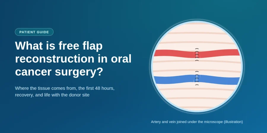
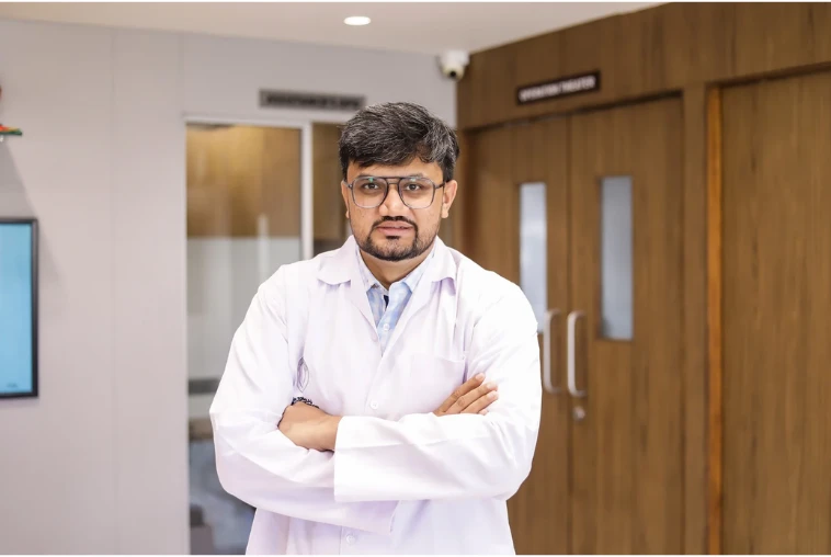
Illustration of an artery and vein joined under a microscope, for a patient guide to free flap reconstruction.

Oral cancer surgery explained

What Is Free Flap Reconstruction in Oral Cancer Surgery?

If your surgeon has said your operation will include "a flap", this guide explains what free flap reconstruction is, where the tissue comes from, what the first days are like, and what your arm or leg will be like afterwards.

Free flap reconstruction is microsurgery that rebuilds the mouth in the same operation that removes the cancer. The surgeon takes a piece of your own living tissue — skin, fat, muscle or bone — from your forearm, thigh or lower leg, along with its own artery and vein. That tissue is moved to the mouth, and the blood vessels are stitched to vessels in the neck under a microscope so the tissue stays alive.

Key takeaways

  • "Free" does not mean no cost. It means the tissue is cut free of its original blood supply, then reconnected to vessels in the neck.
  • The tissue is your own, so your body cannot reject it and you will not need anti-rejection medicine.
  • The defect decides the flap. Soft-tissue gaps take a forearm or thigh flap; a removed jawbone takes the fibula from your lower leg.
  • It happens in one operation, usually six to ten hours, often with two surgical teams working at the same time.
  • The first two days matter most. Nurses check the flap every hour, because almost all problems show up early — and early problems can often be fixed.
  • Losing the fibula does not stop you walking. The lower leg has a second, stronger bone that carries your weight.
  • The point is function — removing a large cancer and still being able to speak, chew and swallow.

What does "free" mean in a free flap?

This word confuses almost everyone. "Free" has nothing to do with money — it describes what happens to the blood supply.

A flap is living tissue moved from one part of the body to another with its blood supply kept intact. In a free flap the surgeon cuts the tissue completely free — artery and vein included — carries it to the mouth, and joins the vessels to vessels in the neck. Vessels this small need an operating microscope, which is why free flap reconstruction is also called microvascular reconstruction.

Surgeons use three different words, and they mean quite different things.

Graft, pedicled flap and free flap — what actually differs

Swipe left to see the full table →

What it is called Does it bring its own blood supply? How it is moved Typically used for
Skin graft No. A thin sheet of skin with no vessels of its own; it must grow new ones from the wound bed. Lifted and laid onto the wound Shallow surface wounds, where nothing underneath is missing
Pedicled flap Yes, but it stays attached — the tissue is swung across while still joined to its original vessels. Rotated from nearby: chest, neck or forehead Medium gaps, and where a free flap is not safe or not available
Free flap Yes, and it is fully detached — its artery and vein are cut and re-joined under a microscope. Carried from a distant site: forearm, thigh, lower leg Large or complex gaps, and anywhere bone or a precise shape is needed

Because the tissue is your own, there is no question of rejection and no anti-rejection medicine.

Diagram comparing a skin graft, a pedicled flap and a free flap by their blood supply.
A graft has no blood supply of its own, a pedicled flap stays attached to its original vessels, and a free flap is detached and re-joined to vessels in the neck.

Why does oral cancer surgery need reconstruction at all?

To cure an oral cancer the surgeon removes the tumour and a rim of healthy tissue around it — the margin, which is what stops the cancer returning at the same spot. On a small, early cancer the gap can be stitched closed. On a larger one it cannot, because pulling the edges of a big hole together does real damage:

  • It tethers the tongue. A tongue stitched to the floor of the mouth cannot move, so speech and swallowing suffer badly.
  • It narrows the mouth. Closing a cheek defect under tension can leave the mouth too tight to open properly.
  • It cannot replace bone. If part of the jaw is gone there is nothing to stitch, and the face loses its shape.

Free flap reconstruction fills the gap with new tissue, so the surgeon never has to choose between removing enough cancer and leaving you able to speak and eat. It is also why the size of a tumour alone does not decide whether surgery is possible — what can be rebuilt afterwards is part of that decision.

Been told your operation will include a flap? Read how Heyansh Oral Cancer Hospital in Ahmedabad plans removal and reconstruction together, as one operation.

Which free flaps are used in oral cancer surgery?

Free flap reconstruction in the mouth relies on a handful of donor sites. Surgeons call these "workhorse" flaps because they are reliable and well understood.

Radial forearm free flap

Taken from the inner forearm near the wrist, the radial forearm free flap gives thin, soft, flexible skin — exactly what the inside of the mouth needs. It is the usual choice for the tongue, floor of the mouth, inner cheek and soft palate, and its long vessels make the microsurgery more straightforward. The trade-off is a visible scar, because the forearm patch is usually covered with a skin graft from the thigh. Your surgeon first tests whether the other wrist artery can supply the whole hand alone.

Fibula free flap

The fibula is the slim outer bone of the lower leg, and the standard choice when jawbone must be rebuilt: long, straight, and strong enough to carry dental implants later. The surgeon cuts small wedges into it to bend it into a jaw curve, and a strip of calf skin can come with it to line the mouth at the same time. Losing it does not stop you walking — the thicker tibia beside it carries almost all your weight.

Anterolateral thigh (ALT) flap

Taken from the outer thigh, the ALT flap provides more bulk than the forearm, so it suits large soft-tissue gaps — a big tongue defect, a full-thickness cheek, or anywhere volume is needed. Its scar is usually closed directly and hidden under clothing.

Less often, surgeons use the scapula (shoulder blade) flap, which carries bone and skin that can be positioned separately; the rectus abdominis flap from the abdominal wall, for very large volumes; or the iliac crest (hip) flap, another bone option for the jaw.

Which free flap will I need?

Patients ask this first, and the honest answer is that you do not choose the flap — the defect does. Once your surgeon knows what must be removed, the options narrow to one or two. Three questions decide it:

  1. Is bone missing, or only soft tissue? Missing bone means a flap that carries bone.
  2. How much bulk does the gap need? A thin lining needs thin tissue; a large hollow needs volume.
  3. Is that donor site safe for you? Circulation, past surgery and old injuries can rule a site out.

How the defect points to the flap. General guidance only — your own plan depends on your scans, your examination and your circulation.

Swipe left to see the full table →

If this was removed Usual first choice Why that one Common alternative
Part of the tongue Radial forearm Thin and flexible, so the remaining tongue can still move freely ALT, if more bulk is needed
Most of the tongue ALT Enough volume to rebuild a mound the tongue can press against to swallow Rectus abdominis
Floor of the mouth Radial forearm Lines the area without tethering the tongue downwards ALT
Inner cheek lining Radial forearm Pliable enough to let the mouth open fully afterwards ALT for full-thickness cheek
Part of the lower jaw Fibula Long, straight bone that can be shaped into a jaw curve and take implants Scapula or iliac crest
Part of the upper jaw Fibula or scapula Rebuilds the bony separation between mouth and nose A prosthetic obturator plate
Jaw plus a lot of soft tissue Scapula Bone and skin can be positioned independently of each other Two flaps in one operation

General patterns described in the surgical literature. Your surgeon's plan may differ for good reasons — ask them to explain it.

Diagram mapping free flap donor sites to the parts of the mouth each one rebuilds.
The three most-used donor sites and the parts of the mouth each one usually rebuilds. General patterns only.

What happens during free flap surgery?

Free flap surgery is one long operation under general anaesthetic, usually six to ten hours. Removal and reconstruction happen in the same sitting — you do not come back later to be rebuilt. Most units run two teams at once: one in the mouth and neck, one raising the flap. Roughly, in order:

  1. The cancer is removed with a margin of healthy tissue. Neck lymph nodes often come out at the same time, which also exposes the vessels the flap will join to.
  2. The flap is raised, still attached to its artery and vein while the surgeon confirms the gap's exact size.
  3. The vessels are divided and the flap detached — from this moment it has no blood supply, so the team works quickly.
  4. The flap is shaped and stitched into the gap. For a jaw, the bone is cut and plated into the right curve first.
  5. The vessels are joined under an operating microscope with stitches finer than a hair. This step — the anastomosis — is the heart of microvascular reconstruction.
  6. The donor site is closed, directly or with a skin graft.
Diagram of two surgical teams working at the same time during free flap reconstruction.
One team removes the cancer while a second raises the flap, so removal and reconstruction happen in a single operation.

Two things are often arranged before you wake, and both are usually temporary. A tracheostomy — a small breathing tube in the neck — keeps your airway clear while the mouth is swollen. A feeding tube through the nose lets the new tissue heal without food passing over it.

The first 48 hours: why the flap is checked every hour

Families are often alarmed that a nurse comes to the bedside every hour through the night, shines a light into the mouth and presses the flap with a gloved finger. This is normal, and it is one of the most important parts of the whole treatment.

The reason is simple. Almost all flap problems appear in the first day or two, and nearly all come down to blood flow through the newly joined vessels. A tiny clot can block the vein or artery. Caught early, the flap can usually be rescued in theatre; missed, the tissue dies. So the staff check a few specific things:

  • Colour — a healthy flap is pink. Pale can mean the artery is blocked; dark blue or purple, the vein.
  • Refill — press the flap and colour should drain and return in a couple of seconds.
  • Warmth and swelling, compared with the tissue around it.
  • A pulse signal — a small handheld Doppler probe listens to blood flowing through the joined vessels.
Illustration of the four checks a nurse makes on a free flap: colour, refill, warmth and Doppler signal.
The four routine checks made on the flap about every hour in the first 24 to 48 hours.

Expect a high-dependency or intensive care bed, no eating by mouth, no speaking if you have a tracheostomy, and a room kept deliberately warm, because cold makes small vessels narrow. None of this means something has gone wrong — it is the standard of care that gives the flap its best chance. Past this window, checks become less frequent and recovery picks up noticeably.

How long is recovery from free flap reconstruction?

Recovery from free flap reconstruction happens in stages. Most people are in hospital for about seven to fourteen days if things go smoothly.

What to expect, stage by stage. Typical timings only — yours will depend on the operation you have and your general health.

Swipe left to see the full table →

Stage Roughly when What is happening
Close monitoring Days 1–2 Hourly flap checks, high-dependency bed, tube feeding, swelling at its worst
Early ward recovery Days 3–7 Sitting up and walking, drains removed, swelling settling, speech and swallow therapy starts
Going home Weeks 1–2 Tracheostomy usually closed, swallowing trials begin, feeding tube out once you can eat enough
Healing and rehabilitation Weeks 4–12 Wounds healed, mouth-opening and swallowing exercises, any radiotherapy begins

Beyond three months improvement continues more gradually, and speech and swallowing usually keep getting better for the best part of a year. Dental implants in a rebuilt jaw are generally considered several months later, once healing is complete and any radiotherapy is finished. Radiotherapy, if needed, usually starts around six weeks after surgery — free flap reconstruction is designed to withstand it, which is another reason tissue with its own blood supply is used rather than a graft.

Speaking and eating after a free flap

This is what most people really want to know, and it deserves an honest answer. Free flap reconstruction restores the gap and the shape of the mouth, but not movement or sensation — the transferred tissue has no muscles under your control and, at first, little feeling.

  • Speech — the flap gives the remaining tongue a firm surface to press against, which is how sounds are formed. Most people are understood again, and therapy makes a real difference to how quickly.
  • Swallowing — relearned in stages, starting with sips and soft food. Keeping a feeding tube for a while as a backup is common and not a sign of failure.
  • Sensation — the flap feels different from normal mouth lining and may stay numb, though some feeling can return over months.
  • Appearance — forearm or thigh skin looks different from the pink lining around it, but settles and becomes less noticeable.
  • Chewing — depends mostly on teeth. If the jaw was rebuilt with a fibula, implants and a denture can restore a great deal later.

Results vary genuinely, depending on how much was removed, which flap was used, whether radiotherapy follows, and how consistently you do the exercises. Ask your surgeon and therapist what is realistic for your own operation.

What happens to the donor site — your arm, leg or thigh?

Most pages about free flap reconstruction skip this. It is usually the second question patients ask, and the first one families ask.

Diagram showing where the donor-site scar sits after a forearm, fibula or thigh free flap.
Typical donor-site scar positions. An illustration, not a patient photograph.

After a radial forearm flap

A patch of inner-forearm skin is taken and usually covered with a thin skin graft from the thigh. The scar is visible, though sleeves cover it. Grip strength is largely preserved. Numbness on the thumb side of the hand is common and may be permanent, and early wrist stiffness improves with physiotherapy.

After a fibula flap

The usual worry is walking, and the answer is reassuring: the fibula is not the weight-bearing bone, and its ends stay in place so the knee and ankle remain stable. You will start walking within days, often with crutches at first, and most people walk normally again within a few weeks. The scar runs down the outer calf. Ankle stiffness, slight weakness lifting the foot and numbness on the outer calf can occur.

After an ALT (thigh) flap

Often the easiest donor site to live with. The wound is usually closed directly, leaving a straight scar down the outer thigh that clothing hides, and thigh strength is generally unaffected because the muscle underneath is left intact. A numb patch on the outer thigh is the most common lasting effect.

Across all three, expect soreness, swelling and stiffness for a few weeks, and expect to do the physiotherapy. The donor site is a real trade-off — but a well-understood one, and it buys back the ability to speak and eat.

Risks and what can go wrong

Free flap reconstruction is major surgery and the risks are real. Modern microsurgery is very reliable and the large majority of free flaps survive, but you should know what the team is watching for.

  • A clot in the joined vessels — the main early risk. Caught quickly, it can often be cleared in theatre and the flap saved.
  • Flap failure — if blood flow cannot be restored the tissue dies and must be removed. Uncommon, and a second reconstruction is then planned.
  • Partial flap loss — only an edge fails and the rest heals, often managed with dressings alone.
  • Infection or wound breakdown, in the mouth or at the donor site.
  • A fistula — a small leak where saliva escapes through the suture line. Usually settles with time.
  • Donor-site problems — a graft not taking, numbness, stiffness or slow healing.
  • General surgical risks — chest infection, blood clots in the legs, and the risks of a long anaesthetic.

Smoking matters more here than almost anywhere else in surgery, because nicotine narrows exactly the small vessels the flap depends on. Stopping beforehand measurably improves your chances, and chewing tobacco, gutkha and similar products should stop completely. Well-controlled diabetes also helps healing.

Who can have free flap reconstruction?

Most people having major oral cancer surgery can. Age alone is not a barrier — studies of microvascular reconstruction in older patients have found it can be done safely, so fitness matters more than the number. Your surgeon will look at:

  • Whether you can tolerate a long anaesthetic — heart and lung health, mainly.
  • Blood supply at the donor site, checked by examination and, where needed, a scan of the vessels.
  • Suitable vessels in the neck — previous surgery or radiotherapy can scar them, though alternatives usually exist.
  • Support at home for the weeks after discharge.

If a free flap is not right for you, that is not the end of reconstruction: a pedicled flap from the chest or neck, or a prosthetic plate, can rebuild a great deal. The right question is not "can I have a free flap" but "what is the best reconstruction for my situation, and why".

One practical point: free flap reconstruction needs specific microsurgical training, an operating microscope and a team able to monitor the flap around the clock, so it is not carried out in every unit. Asking your surgical team whether they perform it, and how often, is a reasonable question.

Questions to ask your surgeon

Take these to your consultation, and bring someone to write the answers down.

  • What exactly are you removing, and how big will the gap be?
  • Which flap are you planning, and why that one for my defect?
  • Where will the donor site be, and what will the scar look like?
  • Will I need a tracheostomy and a feeding tube, and for how long?
  • How long will I be in hospital if all goes well?
  • What should I realistically expect for my speech and swallowing?
  • Will I need radiotherapy afterwards, and could I have dental implants later?
  • How many of these reconstructions does your team do each year?

Want a clear explanation of your own reconstruction plan? Bring your biopsy report and your CT or MRI to Dr Smit A. Desai. He will go through, on a diagram, what has to be removed, which flap it points to and what recovery would look like — or review a plan you have already been given. More about surgery and reconstruction at Heyansh Oral Cancer Hospital, with three clinics in Ahmedabad.

Frequently asked questions

What is free flap reconstruction in oral cancer surgery?
Free flap reconstruction is microsurgery that rebuilds the mouth in the same operation that removes the cancer. The surgeon moves living tissue — skin, fat, muscle or bone — from the forearm, thigh or lower leg, along with its artery and vein, and joins those vessels to neck vessels under a microscope so the tissue stays alive.
Why is it called a "free" flap?
"Free" refers to the blood supply, not the cost. The tissue is cut completely free of its original artery and vein, which are then reconnected to new vessels in the neck. A pedicled flap, by contrast, stays attached to its original blood supply and is swung across from nearby.
What is the difference between a free flap and a graft?
A graft is a thin sheet of tissue with no blood vessels of its own, so it must grow new ones from the wound underneath. A free flap brings its own artery and vein, which is why it can fill a deep gap, carry bone and withstand radiotherapy when a graft cannot.
How long does free flap surgery take?
Usually six to ten hours, sometimes longer for jaw reconstruction. Removing the cancer and rebuilding the mouth happen in one operation, and many units use two surgical teams working at the same time.
How long does it take to recover from free flap surgery?
Most people stay in hospital about seven to fourteen days, and wounds are largely healed by six weeks. Speech and swallowing keep improving for several months with therapy.
Is free flap surgery painful?
Pain after free flap reconstruction is managed with medication and is usually described as soreness and tightness rather than severe pain, in both the mouth and the donor site. Many people find the tubes and swelling harder than the pain.
Will I be able to walk after a fibula free flap?
Yes. The fibula is not the weight-bearing bone — the thicker tibia carries your weight, and the ends of the fibula are left in place to keep the knee and ankle stable. Most people start walking within days and return to walking normally within a few weeks.
What is the success rate of free flap reconstruction?
Modern microvascular reconstruction is highly reliable and the large majority of free flaps survive. Almost all problems appear in the first day or two, which is why the flap is checked hourly at first and why they can often be corrected in theatre.
Can I speak and eat normally after free flap reconstruction?
Most people speak understandably again and return to eating, though both usually change to some degree. The flap restores the shape and surface of the mouth rather than movement, so speech and swallowing therapy are a key part of recovery.
How much does free flap reconstruction cost in India?
Cost depends on the extent of surgery, length of stay, whether intensive care is needed and what treatment follows, so ask for a written estimate for your own plan. Our guide to oral cancer treatment cost in India explains what drives the total.

Would you like a second opinion on your reconstruction plan? Asking for one is a normal part of planning cancer treatment, and surgical teams expect it. Dr Smit A. Desai consults at three clinics in Ahmedabad.

Dr Smit A. Desai, Consultant Oral Cancer Surgeon, Ahmedabad

Medically reviewed by

Dr Smit A. Desai

MDS (Oral & Maxillofacial Surgery) · Consultant Oral Cancer Surgeon

Dr Smit A. Desai is a Consultant Oral Cancer Surgeon practising at Heyansh Oral Cancer Hospital in Ahmedabad, with clinics at Maninagar, Satellite and Nikol. His practice covers oral and head-and-neck cancer surgery, including microvascular free-flap reconstruction performed with an operating microscope, and function-preserving resection of oral cancers. He holds fellowships in oral oncology and in head and neck oncology.

Fellowship in Oral Oncology, AOMSI (2017–19) · Travelling Fellowship in Head & Neck Oncology, FHNO (2018) · MDS (OMFS), CDSRC Ahmedabad · Member of AOMSI, IAOO, IAOMS and FHNO
More about his head and neck cancer practice · Oral cancer specialist in Ahmedabad · About the hospital

About this article

This is general information about free flap reconstruction in oral cancer surgery, not medical advice about your own case. Which reconstruction is right for you depends on your examination, scans and biopsy — always follow your own clinical team's advice.

Sources: StatPearls: Fibula Free Flaps · StatPearls: Radial Forearm Tissue Transfer · Memorial Sloan Kettering: Mandibulectomy and Fibula Free Flap Reconstruction · Katna et al., Clinical outcomes for microvascular reconstruction in oral cancers · Moratin et al., Free-Flap Reconstruction in Early-Stage Oral Cavity Cancer · National Cancer Institute.

Related reading: Oral cancer surgery: what to expect · Signs and symptoms of oral cancer · Tongue cancer surgery in Ahmedabad · Mouth cancer surgery in Ahmedabad · What happens during an oral cancer biopsy · A seven-year survivor's story.

Heyansh Oral Cancer Hospital, Ahmedabad — three clinics: Maninagar (087349 54487) · Satellite (099983 19470) · Nikol / New Naroda (098983 48601). Satellite and Nikol are open Monday to Saturday, 10am–2pm and 5–8pm, and closed on Sunday. Please call the Maninagar clinic for its consulting hours. Contact us or see all branches and contact numbers.

Whales: Giants of the Ocean

Whales are among the largest and most remarkable animals on Earth. These marine mammals live in oceans around the world, from warm tropical waters to the cold seas surrounding the poles.

Life Beneath the Surface

Although whales spend their lives in water, they breathe air through blowholes located on top of their heads. They must regularly return to the surface to breathe before diving again in search of food or traveling through the ocean.

Whales are warm-blooded, give birth to live young, and nurse their calves with milk. A thick layer of fat called blubber helps protect them from cold water and stores energy during long migrations.

Two Main Groups

Whales are generally divided into baleen whales and toothed whales. Baleen whales filter small animals from the water using flexible plates inside their mouths. This group includes blue whales, humpback whales, and gray whales.

Toothed whales use teeth to catch fish, squid, and other prey. Many of them also use echolocation, producing sounds and listening for returning echoes to understand their surroundings. Sperm whales, belugas, and orcas belong to this group.

The Blue Whale

The blue whale is the largest known animal to have ever lived. An adult can grow longer than a city bus and weigh well over one hundred tonnes. Despite its enormous size, it feeds mainly on tiny crustaceans called krill.

Communication and Migration

Whales communicate using clicks, whistles, pulses, and complex songs. Some sounds can travel across great distances underwater. Humpback whales are especially famous for their long, patterned songs.

Many species migrate thousands of kilometres each year. They often feed in cold, nutrient-rich waters before traveling to warmer regions where they mate and give birth.

Protecting Whales

Commercial hunting once caused severe declines in many whale populations. Today, whales also face threats from fishing gear, ship collisions, underwater noise, pollution, and changes to ocean ecosystems.

Conservation programs, safer fishing practices, protected habitats, and international cooperation can help whale populations recover. Protecting whales also supports healthier oceans because these animals play an important role in marine food webs and nutrient cycles.

Whales: Giants of the Ocean

Whales are among the largest and most remarkable animals on Earth. These marine mammals live in oceans around the world, from warm tropical waters to the cold seas surrounding the poles.

Life Beneath the Surface

Although whales spend their lives in water, they breathe air through blowholes located on top of their heads. They must regularly return to the surface to breathe before diving again in search of food or traveling through the ocean.

Whales are warm-blooded, give birth to live young, and nurse their calves with milk. A thick layer of fat called blubber helps protect them from cold water and stores energy during long migrations.

Two Main Groups

Whales are generally divided into baleen whales and toothed whales. Baleen whales filter small animals from the water using flexible plates inside their mouths. This group includes blue whales, humpback whales, and gray whales.

Toothed whales use teeth to catch fish, squid, and other prey. Many of them also use echolocation, producing sounds and listening for returning echoes to understand their surroundings. Sperm whales, belugas, and orcas belong to this group.

The Blue Whale

The blue whale is the largest known animal to have ever lived. An adult can grow longer than a city bus and weigh well over one hundred tonnes. Despite its enormous size, it feeds mainly on tiny crustaceans called krill.

Communication and Migration

Whales communicate using clicks, whistles, pulses, and complex songs. Some sounds can travel across great distances underwater. Humpback whales are especially famous for their long, patterned songs.

Many species migrate thousands of kilometres each year. They often feed in cold, nutrient-rich waters before traveling to warmer regions where they mate and give birth.

Protecting Whales

Commercial hunting once caused severe declines in many whale populations. Today, whales also face threats from fishing gear, ship collisions, underwater noise, pollution, and changes to ocean ecosystems.

Conservation programs, safer fishing practices, protected habitats, and international cooperation can help whale populations recover. Protecting whales also supports healthier oceans because these animals play an important role in marine food webs and nutrient cycles.

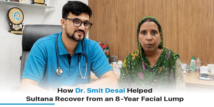
Dr. Smit Desai with Sultana after successful removal of an 8-year facial lump at Heyansh Oral Cancer Hospital.

For eight long years, Sultana lived with a large lump on her face.

She visited multiple doctors hoping someone could help, but almost every consultation ended with the same warning

“The surgery may leave your face permanently disfigured.”

Fear of losing her appearance made her delay treatment while the swelling continued to grow. Some even suspected it could be cancer, leaving her confused and anxious.

Everything changed when she reached Heyansh Oral Cancer Hospital, where Dr. Smit Desai explained the treatment clearly and assured her that the lump could be removed while preserving her facial appearance.

Today, just two months after surgery, she is eating normally, working comfortably, and smiling with confidence again.

Patient Snapshot

Details Information
Patient Sultana
Main Concern Large facial lump for 8 years
Symptoms Large swelling on the face
Previous Experience Multiple consultations with no confidence in surgery
Treatment Surgical removal of facial lump
Surgeon Dr. Smit Desai
Hospital Heyansh Oral Cancer Hospital
Recovery 2 Months
Current Status Eating, working and living normally

When Fear of Facial Disfigurement Delays Treatment

One of the biggest reasons many patients postpone surgery is the fear of facial changes.

That was exactly what happened with Sultana.

During her search for treatment, several doctors warned that removing the lump could affect her facial appearance. Instead of giving her confidence, these consultations increased her anxiety.

As months turned into years, the lump remained untreated.

By the time she reached Dr Smit Desai, she had already spent eight years searching for a solution.

Why Sultana Chose Dr. Smit Desai      

Sultana came to Heyansh Oral Cancer Hospital after someone recommended Dr. Smit Desai .

Unlike her previous consultations, she received detailed counselling about:

  • The possible cause of the lump
  • Surgical planning
  • Expected recovery
  • How facial appearance would be preserved as much as possible

Most importantly, she finally felt confident enough to undergo surgery.

Successfully Removing an 8-Year-Old Facial Lump

The surgical team carefully removed the large facial lump while protecting important facial structures.

The objective wasn’t simply removing the swelling—it was also preserving Sultana’s appearance and helping her return to her normal daily life.

According to her own words, the biggest relief was seeing that her face looked normal after surgery.

Recovery That Restored Her Confidence

Only two months after surgery, Sultana noticed remarkable improvements.

She shared that she could do the following:

  • Eat comfortably
  • Drink normally
  • Perform household work
  • Return to everyday activities
  • Smile confidently

Her recovery exceeded the fears that had stopped her from seeking treatment for years.

What Sultana Says About Her Experience

According to Sultana,

“The doctor explained everything properly and performed the surgery successfully, and my face did not get spoiled.”

She also appreciated the caring environment at Heyansh Oral Cancer Hospital, mentioning that the doctors and staff explained every step patiently and made her feel comfortable throughout her treatment.

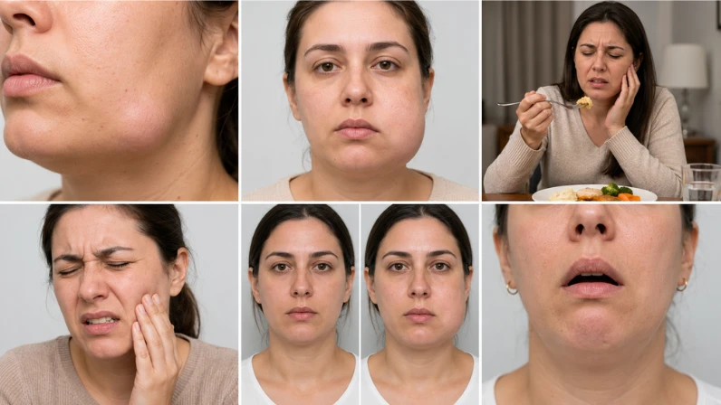
Visual guide showing facial swelling, pain, difficulty eating, changes in appearance, and difficulty opening the mouth.

Signs That Should Never Be Ignored

Not every facial lump is cancer, but a specialist should evaluate every persistent lump.

Seek medical attention if you notice:

  • A lump that continues growing
  • Swelling lasting several weeks
  • Difficulty eating
  • Facial pain
  • Changes in appearance
  • Difficulty opening the mouth

Early diagnosis helps doctors provide better treatment options while reducing complications.

Why Choosing the Right Surgical Team Matters

Treating facial lumps requires experience because surgeons must balance two equally important goals:

  • Removing the disease completely
  • Preserving appearance and normal facial function

This is why patients often seek specialists experienced in head and neck cancer surgery.

Expert Care at Heyansh Oral Cancer Hospital

At Heyansh Oral Cancer Hospital, every patient’s treatment is planned individually.

The hospital focuses on:

  • Oral cancer surgery
  • Facial tumor surgery
  • Head & neck surgery
  • Personalized treatment planning
  • Post-operative rehabilitation
  • Long-term follow-up

More Inspiring Recovery Stories

Every patient’s journey is different.

If you’d like to read more real recovery stories, explore:

Conclusion

Sultana’s story reminds us that delaying treatment because of fear can sometimes make conditions worse.

After living with a facial lump for eight years, she finally found the confidence to undergo surgery under Dr. Smit Desai at Heyansh Oral Cancer Hospital.

Today, she is living proof that expert surgical planning and compassionate care can restore both health and confidence.

If you have a persistent facial swelling or an unexplained lump, don’t ignore it. Consulting an experienced specialist early can make a significant difference in both treatment and recovery.

Frequently Asked Questions

Can a facial lump remain untreated for years?

Yes. Some facial lumps grow slowly over several years, but they should always be examined by a specialist because the cause can vary.

No. Many facial lumps are non-cancerous, but only proper clinical examination and investigations can determine the exact diagnosis.

In many cases, experienced surgeons carefully plan the procedure to preserve facial appearance and important functions whenever possible

Recovery depends on the size and location of the lump, but many patients gradually return to their normal routine within a few weeks to months.

Persistent facial lumps should be evaluated by an experienced head and neck or oral cancer surgeon to determine the appropriate treatment.

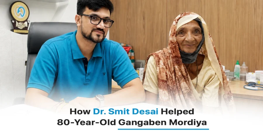
Dr Smit Desai with 80-year-old oral cancer patient Gangaben Mordiya at Heyansh Oral

A Diagnosis That Changed Everything

Gangaben Mordiya is 80 years old. Like most people her age, she just wanted to enjoy her days peacefully with her family. But a small problem in her mouth slowly turned into a diagnosis that changed everything: oral cancer.

At first, it did not seem like anything serious. But as the days passed, the symptoms grew harder to ignore, and worry started creeping into the family’s mind. What followed was fear, confusion, and a lot of questions about what would happen next.

That is when the family decided to visit Heyansh Oral Cancer Hospital, known as one of the best oral cancer hospitals for treating cases like this, and met Dr. Smit Desai, who would go on to guide Gangaben through her entire treatment journey.

Today, Gangaben is healthy, smiling, and back to her normal life. This is the story of how that happened.

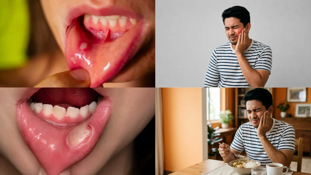
Oral cancer symptoms including mouth ulcer, jaw pain, and eating discomfort shown with patient awareness visuals

Understanding the Problem: The Symptoms Gangaben Could Not Ignore

Patient Snapshot

  • Patient: Gangaben Mordiya
  • Age: 80 Years
  • Diagnosis: Oral Cancer
  • Treatment: Oral Cancer Surgery
  • Specialist: Dr. Smit Desai
  • Hospital: Heyansh Oral Cancer Hospital
  • Current Status: Living a healthy life with no reported recurrence

It started small. A little discomfort in the mouth. Maybe a small ulcer that did not seem like a big deal. But over time, things got worse.

She began to find it hard to eat properly. Talking became uncomfortable. Simple daily activities that she never had to think about before, like chewing food or speaking clearly, started to feel like a struggle.

This is exactly the kind of warning sign that should never be ignored, especially:

  • A mouth ulcer that does not heal after a couple of weeks
  • Ongoing pain inside the mouth
  • Swelling that does not go down
  • Trouble eating or speaking normally

Once the family saw that these symptoms were not going away on their own, they knew it was time to see a specialist instead of waiting any longer. That decision led them straight to Heyansh Oral Cancer Hospital, a hospital built specifically to treat oral cancer cases like Gangaben’s.

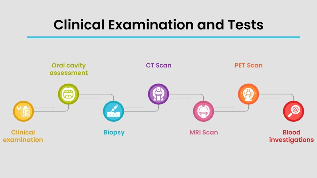
Oral cancer diagnosis process showing clinical examination, biopsy, CT scan, MRI scan, PET scan, and blood investigations

The Diagnosis: Finding the Root Cause

Clinical Examination and Tests

Once Gangaben reached the hospital, Dr. Smit Desai began with a detailed examination of her mouth to understand exactly what was going on. This was not a quick look; it was a thorough check to study the affected area closely.

To confirm what was suspected, a biopsy was done. In simple words, this means a small sample of tissue was taken from the affected area and tested in a lab to check whether it was actually cancer.

Along with that, CT and MRI scans were carried out. These scans work like detailed internal pictures, helping the doctor see the exact size of the tumor and where it was located. The team also checked her nearby lymph nodes, which are small glands in the body that can show if the cancer has started spreading, and this helped them understand the overall stage of her cancer.

Why This Case Needed Special Attention

Treating oral cancer is already a delicate task, but treating it in an 80-year-old patient comes with its own extra layer of care. The body does not heal as quickly at this age, and there are usually other health factors to think about as well.

This meant Dr. Smit Desai and his team at Heyansh Oral Cancer Hospital had to carefully balance two things at once: removing the cancer completely and making sure Gangaben’s overall health and recovery were never put at risk.

Creating a Treatment Plan: Looking Beyond Age

Dr. smit Desai’s Approach

Before deciding on any treatment, Dr. Smit Desai made sure to fully evaluate Gangaben’s overall health and whether she was fit for surgery. Age was never treated as something that could simply rule out treatment; instead, it was treated as a factor to plan carefully around.

The treatment options were discussed openly with Gangaben and her family, so they understood exactly what to expect at each step. The main goal was clear from the start: remove the cancer completely while also protecting important everyday functions like her ability to speak and swallow comfortably.

This kind of personalized planning is exactly why so many families trust Heyansh Oral Cancer Hospital, often regarded as the best oral cancer hospital for handling complex cases with the right balance of safety and results.

The Surgery: Removing the Cancer Safely

How the Oral Cancer Was Treated

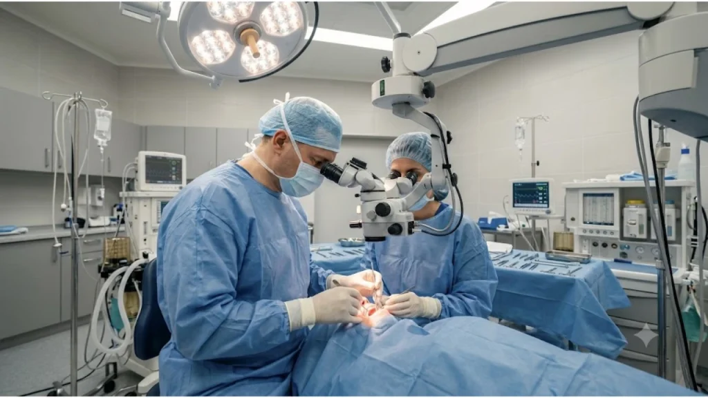
The treatment moved forward with a procedure called a wide local excision. In simple terms, this means the tumor was removed along with a safe margin of healthy tissue around it to make sure no cancer cells were left behind.

Wherever needed, the team also carefully checked and managed nearby lymph nodes, since this plays an important role in preventing the cancer from coming back. Every step of the surgery was planned not just to remove the cancer, but also to help Gangaben maintain a good quality of life afterward.

Managing Surgery at 80 Years of Age

Performing surgery on an 80-year-old patient required extra precaution at every stage.

Pre-Operative Medical Assessment

Gangaben’s overall health was checked thoroughly before surgery to confirm she was strong enough for the procedure.

Specialized Anesthesia Planning

Extra Safety Measures During SurgeryThe anaesthesia was planned carefully, keeping her age and health in mind, to keep her safe throughout the operation.

The surgical team stayed alert to any age-related risks throughout the procedure.

Close Post-Operative Monitoring

Right after surgery, she was watched closely to catch any complications early and support smooth healing.

This careful, age-aware approach is one of the reasons Dr. Smit Desai and Heyansh Oral Cancer Hospital are trusted by so many families dealing with oral cancer in elderly loved ones.

Parameter Details
Surgery Duration 2–3 Hours
Tumor Removed Yes
Reconstruction No
ICU Stay 1 Day (overnight observation)
Hospital Stay 5–6 Days

Step-by-Step: Gangaben’s Treatment Journey

Here is a clear, step-by-step look at how Gangaben’s treatment unfolded at Heyansh Oral Cancer Hospital, from the first symptom to full recovery.

Phase 1: Diagnosis

Step 1: Noticing the Symptoms

Persistent mouth discomfort and difficulty eating led the family to seek medical help.

Step 2: First Visit to Heyansh Oral Cancer Hospital

Gangaben was examined by Dr. Smit Desai, who began assessing her condition.

Step 3: Biopsy

A small tissue sample was taken and tested to confirm oral cancer.

Step 4: CT/MRI Scans

Imaging tests were done to study the size, location, and spread of the tumor.

Step 5: Lymph Node and Staging Assessment

The team checked nearby lymph nodes to understand the stage of the cancer.

Phase 2: Planning

Step 6: Health and Fitness Evaluation

Gangaben’s overall health was reviewed to confirm she was fit for surgery at 80 years of age.

Step 7: Treatment Planning

Dr. Smit Desai designed a treatment plan focused on completely removing the cancer while protecting her speech and swallowing ability.

Phase 3: Surgery

Step 8: Pre-Operative Preparation

Medical checks and anaesthesia planning were completed before surgery.

Step 9: Wide Local Excision Surgery

Dr. Smit Desai performed the surgery, removing the tumour along with a safe margin of healthy tissue.

Phase 4: Recovery

Step 10: Post-Operative Care

Gangaben was closely monitored for pain management, healing, and any early complications.

Step 11: Recovery and Nutritional Support

She was gradually guided back to eating and speaking comfortably.

Step 12: Follow-Up and Rehabilitation

Regular check-ups, oral examinations, and cancer surveillance were scheduled to track her progress and catch any issues early.

This clear, step-by-step process at Heyansh Oral Cancer Hospital ensured that nothing was rushed, and every stage of Gangaben’s treatment was handled with care.

Recovery After Surgery: The Road Back to Normal Life

The First Few Weeks

In the weeks right after surgery, the focus was on healing. The team at Heyansh Oral Cancer Hospital kept a close watch on her recovery, managed her pain, and made sure she was getting the right nutrition to support her body’s healing process.

Slowly but surely, Gangaben began eating and speaking more comfortably again, something her family had been hoping for since the very beginning of this journey.

Follow-Up and Rehabilitation

Recovery did not stop once she left the hospital. Regular check-ups were scheduled so Dr. Smit Desai could continue monitoring her healing. Ongoing oral examinations and cancer surveillance were part of this routine, helping the team catch any potential issues early.

She was also guided on how to maintain good oral health going forward, to lower the chances of any future complications.

The Outcome: A New Chapter for Gangaben

Life Before Treatment

Before treatment, life was difficult for Gangaben. There was pain, discomfort, and a lot of uncertainty. Everyday things like eating and talking, things most of us never think twice about, had become a real struggle.

Life After Treatment

After her surgery and recovery at Heyansh Oral Cancer Hospital, everything changed for the better:

  • A successful recovery after her oral cancer surgery
  • A noticeably improved quality of life
  • The ability to return to her normal daily routine
  • No reported recurrence, along with continued follow-up care to keep it that way

Today, at 80 years old, Gangaben is living proof that the right treatment and the right team can truly turn things around.

Before Treatment After Treatment
Pain while eating Comfortable eating
Non-healing ulcer Healed surgical site
Difficulty swallowing Improved swallowing
Reduced nutrition Better nutritional intake

What This Case Teaches Us About Oral Cancer

Symptoms That Should Never Be Ignored

Gangaben’s story is a reminder to pay attention to these warning signs:

  • Mouth ulcers that do not heal
  • Persistent pain inside the mouth
  • Difficulty swallowing
  • White or red patches inside the mouth
  • Swelling that does not go away

Why Early Treatment Matters

The earlier oral cancer is caught, the better things usually turn out. Early treatment generally means:

  • Better overall outcomes
  • Less extensive surgery
  • A higher chance of long-term recovery

Dr. Smit Desai’s Message to Patients and Families

According to Dr. Smit Desai, Gangaben’s case is proof that oral cancer can often be treated successfully, even in elderly patients. Age alone should never be a reason to avoid seeking treatment.

His message is simple: early diagnosis combined with expert surgical care, the kind provided at Heyansh Oral Cancer Hospital, can make a real and lasting difference in a patient’s life.

Conclusion: Gangaben’s Journey Is a Story of Hope

Gangaben Mordiya’s journey, from the first worrying symptoms to a successful surgery and a happy recovery, is a powerful reminder of what is possible with the right care. With Dr. Smit Desai guiding her treatment and the dedicated team at Heyansh Oral Cancer Hospital supporting her every step of the way, she now enjoys a healthy, active life at 80 years old.

If you or someone you love is experiencing symptoms like persistent mouth ulcers, pain, or swelling, do not wait. Reach out to Heyansh Oral Cancer Hospital, widely regarded as the best oral cancer hospital for these cases, and let Dr. Smit Desai and his team guide you toward the same kind of hopeful outcome that Gangaben experienced.

Frequently Asked Questions

A wide local excision removes the tumor along with a margin of healthy tissue around it. It was chosen for Gangaben because it lowers the risk of leftover cancer cells and reduces the chance of recurrence.

A biopsy checks tissue cells under a microscope for cancerous changes, something a visual exam alone cannot confirm. This is why Dr. Smith Patel ordered one before starting any treatment plan.

A biopsy confirms whether cancer is present, but CT and MRI scans show its exact size, location, and depth. This information is needed to plan the surgery and check if nearby structures are affected.

Yes. If lymph nodes show signs of cancer spread, treatment may need to include lymph node removal or additional therapy alongside surgery, which is why staging is checked before planning treatment.

It carries more risk than in younger patients, which is why a specialized anesthesia plan and a full pre-operative health assessment were done before Gangaben's surgery to keep her safe.

Recovery timelines vary, but most patients are guided through a gradual return to eating, starting with soft foods and nutritional support, before slowly moving back to a normal diet under medical supervision.

It involves scheduled follow-up visits, oral examinations, and sometimes imaging to check the surgical site and surrounding tissue for any early signs of recurrence.

Recurrence is possible with any cancer surgery, which is why regular follow-up and surveillance, like the kind Gangaben continues to receive, remain an important part of long-term care.

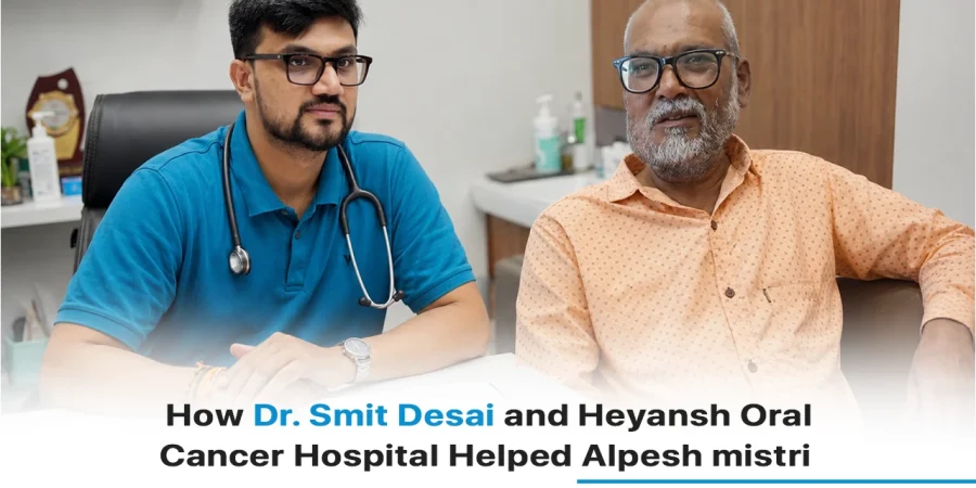
Dr Smit Desai with oral cancer patient Alpesh Mistri at Heyansh Oral Cancer Hospital after successful treatment and recovery.

Patient Snapshot

Detail Information
Patient Alpesh Mistri
Age 63 years
Diagnosis Oral Cancer
Treatment Oral Cancer Surgery
Specialist Dr. Smit Desai
Hospital Heyansh Oral Cancer Hospital
Time Since Treatment 7 Years
Estimated Recovery Time 4–6 Weeks for initial healing
Current Status Eating normally, living happily, no reported recurrence

When Oral Cancer Threatened to Change Everything

Hearing the word “cancer” can change everything in a single moment. That is exactly what happened to this patient when he was told he had oral cancer. Like most people, he had a first reaction of fear. Fear of surgery, fear of pain, and fear of not knowing what life would look like afterwards.

But with timely treatment at Heyansh Oral Cancer Hospital, guided by Dr. Smit Desai, he was able to begin a completely new chapter in his life. Today, 7 years after his treatment, he eats normally, feels confident, and enjoys life just like before, maybe even more.

This is his story, a simple reminder of how the right treatment, the right hospital, and the right mindset can truly change everything.

Oral cancer warning signs including mouth ulcer, jaw pain, chewing discomfort, and difficulty eating

The Early Warning Signs That Led to a Diagnosis

Symptoms That Could Not Be Ignored

It usually starts small. For this patient, it began with a few signs that slowly got harder to ignore:

  • A mouth ulcer that simply would not heal
  • Pain while chewing food
  • Trouble eating certain foods comfortably
  • A burning feeling or general discomfort in the mouth
  • A gradual drop in how much and how comfortably he could eat

At first, these signs might seem minor. But when they do not go away after a couple of weeks, they are the body’s way of asking for attention.

Seeking Specialized Care

As the symptoms continued, his family grew more worried. Eating had become uncomfortable, and something clearly was not right. That worry led to an important decision: it was time to see a specialist instead of waiting any longer.

This decision brought him to Heyansh Oral Cancer Hospital, widely known as the best oral cancer hospital for patients dealing with exactly this kind of problem.

Understanding the Diagnosis

Comprehensive Evaluation and Cancer Detection

Once he arrived at Heyansh Oral Cancer Hospital, Dr. Smit Desai began a complete evaluation to understand what was really going on. This included:

  • A detailed examination of the inside of his mouth
  • A biopsy, where a small piece of tissue is removed and tested in a lab to confirm oral cancer
  • Imaging studies like CT or MRI scans, which work like detailed internal pictures to show the size and location of the tumor
  • A check of nearby lymph nodes to see if the cancer had spread
  • Cancer staging, which simply means understanding how advanced the cancer was, so the right treatment plan could be created

Why Early Diagnosis Matters

Catching oral cancer early makes a real difference. It usually means:

  • Better treatment outcomes overall
  • A higher chance of removing the cancer completely
  • A better chance of keeping important functions like speech and swallowing working normally

This is exactly why Dr. Smit Desai always stresses that patients should never delay a check-up when something feels wrong in the mouth.

Building a Personalized Treatment Plan

More Than Just Treating Cancer

For Dr. Smit Desai and the team at Heyansh Oral Cancer Hospital, treatment was never just about removing cancer cells. It was about looking at the patient as a whole person. This meant:

  • Understanding his overall health condition
  • Checking his nutritional status, since eating difficulties can affect overall strength
  • Planning treatment in a way that balanced controlling the cancer with protecting his everyday quality of life

Treatment Goals

The treatment plan was built around four simple goals:

  1. Remove the cancer completely
  2. Preserve normal oral function, like eating and speaking
  3. Help him regain his confidence and independence
  4. Reduce the risk of the cancer coming back

The Treatment Journey

Surgical Management of Oral Cancer

Like many oral cancer cases handled at Heyansh Oral Cancer Hospital, his treatment plan included:

  • A wide local excision, where the cancerous tissue is removed along with a safe margin of healthy tissue around it
  • Careful surgical margins to lower the risk of recurrence
  • Assessment and treatment of nearby lymph nodes, wherever necessary

Recovery and Rehabilitation

After the surgery, the focus shifted fully toward healing and getting back to normal life. This included:

  • Healing of the surgical area under close observation
  • Nutritional support to rebuild his strength
  • Gradually regaining comfortable eating habits
  • Support for speech and swallowing recovery
  • Regular follow-up evaluations to track his progress
Step-by-step oral cancer treatment process showing symptoms, diagnosis, biopsy, scans, surgery, recovery, and follow-up care

Step-by-Step: How His Treatment Unfolded

Here is a simple breakdown of his treatment journey at Heyansh Oral Cancer Hospital:

Step 1: Noticing the Symptoms

Persistent mouth ulcers, pain while chewing, and discomfort while eating became impossible to ignore.

Step 2: Visiting Heyansh Oral Cancer Hospital

The family decided to consult a specialist, leading them to Dr. Smit Desai at Heyansh Oral Cancer Hospital.

Step 3: Detailed Oral Examination

Dr. Smit Desai carefully examined the affected area to understand the problem.

Step 4: Biopsy

A small tissue sample was tested to confirm that it was oral cancer.

Step 5: CT/MRI Imaging

Scans were done to check the size, location, and spread of the tumour.

Step 6: Lymph Node Assessment and Staging

Nearby lymph nodes were checked, and the cancer was staged to guide the treatment plan.

Step 7: Treatment Planning

Dr. Smit Desai designed a personalised plan focused on removing the cancer while protecting his speech and swallowing ability.

Step 8: Wide Local Excision Surgery

The surgery was performed to remove the tumour along with a safe margin of healthy tissue.

Step 9: Post-Operative Care

He was monitored closely for healing, pain control, and early signs of any complications.

Step 10: Nutritional and Speech Rehabilitation

Support was provided to help him return to comfortable eating and clear speech.

Step 11: Long-Term Follow-Up

Regular check-ups at Heyansh Oral Cancer Hospital continued for years to monitor his recovery and catch any issues early.

This structured, step-by-step approach is exactly why so many patients trust Dr. Smit Desai and consider Heyansh Oral Cancer Hospital the best oral cancer hospital for this kind of treatment.

The Power of Positivity During Recovery

Healing Beyond Surgery

Surgery was only one part of his recovery. The rest came from something just as powerful:

  • A positive attitude
  • Strong determination
  • Support from his family
  • Staying consistent with follow-up care

As he shared in his own testimonial, completing treatment along with strong willpower gave him a “new life”.

Seven Years Later: Living Life Normally Again

Returning to Everyday Activities

After completing his treatment at Heyansh Oral Cancer Hospital, life slowly returned to normal:

  • Eating normally again, without pain or discomfort
  • Improved comfort and confidence in daily activities
  • The ability to truly enjoy daily life again
  • Staying consistent with regular health check-ups

A Long-Term Success Story

Seven years after his treatment, he continues to live a healthy and active life. His story is a clear example of how effective treatment under Dr. Smit Desai, combined with proper follow-up, can lead to excellent long-term outcomes.

What This Case Teaches Us About Oral Cancer

Symptoms You Should Never Ignore

  • Mouth ulcers lasting more than two weeks
  • Persistent oral pain
  • Difficulty swallowing
  • White or red patches inside the mouth
  • Unexplained bleeding or swelling

Why Specialized Oral Cancer Care Matters

  • Accurate diagnosis from the very first visit
  • Expert surgical planning
  • Protecting important everyday functions
  • Long-term monitoring and ongoing support

This is exactly the kind of care that Heyansh Oral Cancer Hospital is known for, which is why it is often called the best oral cancer hospital by patients and families alike.

The Role of Long-Term Follow-Up in Oral Cancer Recovery

Life After Cancer Treatment

Even after a successful surgery, regular follow-up remains important. At Heyansh Oral Cancer Hospital, this means:

  • Monitoring for any signs of recurrence
  • Assessing how well oral function has recovered
  • Maintaining good nutritional health
  • Making sure the quality of life stays high for years to come

The Importance of Annual Reviews

Yearly check-ups with Dr. Smit Desai help catch any small issues early, long before they could become serious problems. This is one of the biggest reasons patients remain healthy for years after treatment.

A Message of Hope for Oral Cancer Patients

“Treatment, Positive Attitude, and Willpower Can Give a New Life”

This patient’s journey is proof that oral cancer treatment is not just about removing disease. It is also about helping people get their confidence, independence, and happiness back. Seven years after his treatment at Heyansh Oral Cancer Hospital, his ability to eat normally and live joyfully stands as real proof of what is possible with expert care from Dr. Smit Desai and a determined mindset.

Conclusion: A Journey From Cancer to Confidence

From the first warning signs to a successful surgery and seven happy years of recovery, this patient’s story shows just how much is possible with the right care. With Dr. Smit Desai guiding his treatment and Heyansh Oral Cancer Hospital supporting him at every step, he now lives a healthy, confident, and joyful life.

If you or a loved one is noticing symptoms like mouth ulcers, pain, or difficulty eating, do not wait. Reach out to Heyansh Oral Cancer Hospital, regarded as the best oral cancer hospital for cases like this, and let Dr. Smit Desai and his team help you write your own story of recovery and hope.

Frequently Asked Questions

Initial healing generally takes around 4 to 6 weeks, though full recovery of comfortable eating and speech can take a bit longer depending on the individual.

Scans show the size and location of a growth, but only a biopsy can confirm under a microscope whether it is actually cancerous.

No treatment can guarantee zero risk, which is why regular follow-up and annual reviews remain an important part of staying cancer-free long-term.

Lymph nodes can show early signs of cancer spread that are not visible from the tumour alone, so checking them helps plan the right level of treatment.

Yes, with proper rehabilitation and nutritional support, most patients gradually return to eating normally, as seen in this patient's 7-year recovery.

Most specialists, including Dr. Smith Patel, recommend regular check-ups, often annually, once a patient is well past the initial recovery period, to catch any issues early.