Oral cancer surgery explained
What Is Free Flap Reconstruction in Oral Cancer Surgery?
If your surgeon has said your operation will include "a flap", this guide explains what free flap reconstruction is, where the tissue comes from, what the first days are like, and what your arm or leg will be like afterwards.
 Medically reviewed by Dr Smit A. Desai, MDS (OMFS) — Consultant Oral Cancer Surgeon
Published 28 October 2026 · Last reviewed 28 October 2026 · About 12 minutes to read
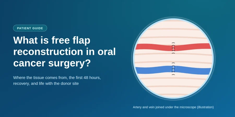
Free flap reconstruction is microsurgery that rebuilds the mouth in the same operation that removes the cancer. The surgeon takes a piece of your own living tissue — skin, fat, muscle or bone — from your forearm, thigh or lower leg, along with its own artery and vein. That tissue is moved to the mouth, and the blood vessels are stitched to vessels in the neck under a microscope so the tissue stays alive.
Key takeaways
- "Free" does not mean no cost. It means the tissue is cut free of its original blood supply, then reconnected to vessels in the neck.
- The tissue is your own, so your body cannot reject it and you will not need anti-rejection medicine.
- The defect decides the flap. Soft-tissue gaps take a forearm or thigh flap; a removed jawbone takes the fibula from your lower leg.
- It happens in one operation, usually six to ten hours, often with two surgical teams working at the same time.
- The first two days matter most. Nurses check the flap every hour, because almost all problems show up early — and early problems can often be fixed.
- Losing the fibula does not stop you walking. The lower leg has a second, stronger bone that carries your weight.
- The point is function — removing a large cancer and still being able to speak, chew and swallow.
What does "free" mean in a free flap?
This word confuses almost everyone. "Free" has nothing to do with money — it describes what happens to the blood supply.
A flap is living tissue moved from one part of the body to another with its blood supply kept intact. In a free flap the surgeon cuts the tissue completely free — artery and vein included — carries it to the mouth, and joins the vessels to vessels in the neck. Vessels this small need an operating microscope, which is why free flap reconstruction is also called microvascular reconstruction.
Surgeons use three different words, and they mean quite different things.
Graft, pedicled flap and free flap — what actually differs
Swipe left to see the full table →
| What it is called | Does it bring its own blood supply? | How it is moved | Typically used for |
|---|---|---|---|
| Skin graft | No. A thin sheet of skin with no vessels of its own; it must grow new ones from the wound bed. | Lifted and laid onto the wound | Shallow surface wounds, where nothing underneath is missing |
| Pedicled flap | Yes, but it stays attached — the tissue is swung across while still joined to its original vessels. | Rotated from nearby: chest, neck or forehead | Medium gaps, and where a free flap is not safe or not available |
| Free flap | Yes, and it is fully detached — its artery and vein are cut and re-joined under a microscope. | Carried from a distant site: forearm, thigh, lower leg | Large or complex gaps, and anywhere bone or a precise shape is needed |
Because the tissue is your own, there is no question of rejection and no anti-rejection medicine.

Why does oral cancer surgery need reconstruction at all?
To cure an oral cancer the surgeon removes the tumour and a rim of healthy tissue around it — the margin, which is what stops the cancer returning at the same spot. On a small, early cancer the gap can be stitched closed. On a larger one it cannot, because pulling the edges of a big hole together does real damage:
- It tethers the tongue. A tongue stitched to the floor of the mouth cannot move, so speech and swallowing suffer badly.
- It narrows the mouth. Closing a cheek defect under tension can leave the mouth too tight to open properly.
- It cannot replace bone. If part of the jaw is gone there is nothing to stitch, and the face loses its shape.
Free flap reconstruction fills the gap with new tissue, so the surgeon never has to choose between removing enough cancer and leaving you able to speak and eat. It is also why the size of a tumour alone does not decide whether surgery is possible — what can be rebuilt afterwards is part of that decision.
Been told your operation will include a flap? Read how Heyansh Oral Cancer Hospital in Ahmedabad plans removal and reconstruction together, as one operation.
Which free flaps are used in oral cancer surgery?
Free flap reconstruction in the mouth relies on a handful of donor sites. Surgeons call these "workhorse" flaps because they are reliable and well understood.
Radial forearm free flap
Taken from the inner forearm near the wrist, the radial forearm free flap gives thin, soft, flexible skin — exactly what the inside of the mouth needs. It is the usual choice for the tongue, floor of the mouth, inner cheek and soft palate, and its long vessels make the microsurgery more straightforward. The trade-off is a visible scar, because the forearm patch is usually covered with a skin graft from the thigh. Your surgeon first tests whether the other wrist artery can supply the whole hand alone.
Fibula free flap
The fibula is the slim outer bone of the lower leg, and the standard choice when jawbone must be rebuilt: long, straight, and strong enough to carry dental implants later. The surgeon cuts small wedges into it to bend it into a jaw curve, and a strip of calf skin can come with it to line the mouth at the same time. Losing it does not stop you walking — the thicker tibia beside it carries almost all your weight.
Anterolateral thigh (ALT) flap
Taken from the outer thigh, the ALT flap provides more bulk than the forearm, so it suits large soft-tissue gaps — a big tongue defect, a full-thickness cheek, or anywhere volume is needed. Its scar is usually closed directly and hidden under clothing.
Less often, surgeons use the scapula (shoulder blade) flap, which carries bone and skin that can be positioned separately; the rectus abdominis flap from the abdominal wall, for very large volumes; or the iliac crest (hip) flap, another bone option for the jaw.
Which free flap will I need?
Patients ask this first, and the honest answer is that you do not choose the flap — the defect does. Once your surgeon knows what must be removed, the options narrow to one or two. Three questions decide it:
- Is bone missing, or only soft tissue? Missing bone means a flap that carries bone.
- How much bulk does the gap need? A thin lining needs thin tissue; a large hollow needs volume.
- Is that donor site safe for you? Circulation, past surgery and old injuries can rule a site out.
How the defect points to the flap. General guidance only — your own plan depends on your scans, your examination and your circulation.
Swipe left to see the full table →
| If this was removed | Usual first choice | Why that one | Common alternative |
|---|---|---|---|
| Part of the tongue | Radial forearm | Thin and flexible, so the remaining tongue can still move freely | ALT, if more bulk is needed |
| Most of the tongue | ALT | Enough volume to rebuild a mound the tongue can press against to swallow | Rectus abdominis |
| Floor of the mouth | Radial forearm | Lines the area without tethering the tongue downwards | ALT |
| Inner cheek lining | Radial forearm | Pliable enough to let the mouth open fully afterwards | ALT for full-thickness cheek |
| Part of the lower jaw | Fibula | Long, straight bone that can be shaped into a jaw curve and take implants | Scapula or iliac crest |
| Part of the upper jaw | Fibula or scapula | Rebuilds the bony separation between mouth and nose | A prosthetic obturator plate |
| Jaw plus a lot of soft tissue | Scapula | Bone and skin can be positioned independently of each other | Two flaps in one operation |
General patterns described in the surgical literature. Your surgeon's plan may differ for good reasons — ask them to explain it.

What happens during free flap surgery?
Free flap surgery is one long operation under general anaesthetic, usually six to ten hours. Removal and reconstruction happen in the same sitting — you do not come back later to be rebuilt. Most units run two teams at once: one in the mouth and neck, one raising the flap. Roughly, in order:
- The cancer is removed with a margin of healthy tissue. Neck lymph nodes often come out at the same time, which also exposes the vessels the flap will join to.
- The flap is raised, still attached to its artery and vein while the surgeon confirms the gap's exact size.
- The vessels are divided and the flap detached — from this moment it has no blood supply, so the team works quickly.
- The flap is shaped and stitched into the gap. For a jaw, the bone is cut and plated into the right curve first.
- The vessels are joined under an operating microscope with stitches finer than a hair. This step — the anastomosis — is the heart of microvascular reconstruction.
- The donor site is closed, directly or with a skin graft.

Two things are often arranged before you wake, and both are usually temporary. A tracheostomy — a small breathing tube in the neck — keeps your airway clear while the mouth is swollen. A feeding tube through the nose lets the new tissue heal without food passing over it.
The first 48 hours: why the flap is checked every hour
Families are often alarmed that a nurse comes to the bedside every hour through the night, shines a light into the mouth and presses the flap with a gloved finger. This is normal, and it is one of the most important parts of the whole treatment.
The reason is simple. Almost all flap problems appear in the first day or two, and nearly all come down to blood flow through the newly joined vessels. A tiny clot can block the vein or artery. Caught early, the flap can usually be rescued in theatre; missed, the tissue dies. So the staff check a few specific things:
- Colour — a healthy flap is pink. Pale can mean the artery is blocked; dark blue or purple, the vein.
- Refill — press the flap and colour should drain and return in a couple of seconds.
- Warmth and swelling, compared with the tissue around it.
- A pulse signal — a small handheld Doppler probe listens to blood flowing through the joined vessels.

Expect a high-dependency or intensive care bed, no eating by mouth, no speaking if you have a tracheostomy, and a room kept deliberately warm, because cold makes small vessels narrow. None of this means something has gone wrong — it is the standard of care that gives the flap its best chance. Past this window, checks become less frequent and recovery picks up noticeably.
How long is recovery from free flap reconstruction?
Recovery from free flap reconstruction happens in stages. Most people are in hospital for about seven to fourteen days if things go smoothly.
What to expect, stage by stage. Typical timings only — yours will depend on the operation you have and your general health.
Swipe left to see the full table →
| Stage | Roughly when | What is happening |
|---|---|---|
| Close monitoring | Days 1–2 | Hourly flap checks, high-dependency bed, tube feeding, swelling at its worst |
| Early ward recovery | Days 3–7 | Sitting up and walking, drains removed, swelling settling, speech and swallow therapy starts |
| Going home | Weeks 1–2 | Tracheostomy usually closed, swallowing trials begin, feeding tube out once you can eat enough |
| Healing and rehabilitation | Weeks 4–12 | Wounds healed, mouth-opening and swallowing exercises, any radiotherapy begins |
Beyond three months improvement continues more gradually, and speech and swallowing usually keep getting better for the best part of a year. Dental implants in a rebuilt jaw are generally considered several months later, once healing is complete and any radiotherapy is finished. Radiotherapy, if needed, usually starts around six weeks after surgery — free flap reconstruction is designed to withstand it, which is another reason tissue with its own blood supply is used rather than a graft.
Speaking and eating after a free flap
This is what most people really want to know, and it deserves an honest answer. Free flap reconstruction restores the gap and the shape of the mouth, but not movement or sensation — the transferred tissue has no muscles under your control and, at first, little feeling.
- Speech — the flap gives the remaining tongue a firm surface to press against, which is how sounds are formed. Most people are understood again, and therapy makes a real difference to how quickly.
- Swallowing — relearned in stages, starting with sips and soft food. Keeping a feeding tube for a while as a backup is common and not a sign of failure.
- Sensation — the flap feels different from normal mouth lining and may stay numb, though some feeling can return over months.
- Appearance — forearm or thigh skin looks different from the pink lining around it, but settles and becomes less noticeable.
- Chewing — depends mostly on teeth. If the jaw was rebuilt with a fibula, implants and a denture can restore a great deal later.
Results vary genuinely, depending on how much was removed, which flap was used, whether radiotherapy follows, and how consistently you do the exercises. Ask your surgeon and therapist what is realistic for your own operation.
What happens to the donor site — your arm, leg or thigh?
Most pages about free flap reconstruction skip this. It is usually the second question patients ask, and the first one families ask.

After a radial forearm flap
A patch of inner-forearm skin is taken and usually covered with a thin skin graft from the thigh. The scar is visible, though sleeves cover it. Grip strength is largely preserved. Numbness on the thumb side of the hand is common and may be permanent, and early wrist stiffness improves with physiotherapy.
After a fibula flap
The usual worry is walking, and the answer is reassuring: the fibula is not the weight-bearing bone, and its ends stay in place so the knee and ankle remain stable. You will start walking within days, often with crutches at first, and most people walk normally again within a few weeks. The scar runs down the outer calf. Ankle stiffness, slight weakness lifting the foot and numbness on the outer calf can occur.
After an ALT (thigh) flap
Often the easiest donor site to live with. The wound is usually closed directly, leaving a straight scar down the outer thigh that clothing hides, and thigh strength is generally unaffected because the muscle underneath is left intact. A numb patch on the outer thigh is the most common lasting effect.
Across all three, expect soreness, swelling and stiffness for a few weeks, and expect to do the physiotherapy. The donor site is a real trade-off — but a well-understood one, and it buys back the ability to speak and eat.
Risks and what can go wrong
Free flap reconstruction is major surgery and the risks are real. Modern microsurgery is very reliable and the large majority of free flaps survive, but you should know what the team is watching for.
- A clot in the joined vessels — the main early risk. Caught quickly, it can often be cleared in theatre and the flap saved.
- Flap failure — if blood flow cannot be restored the tissue dies and must be removed. Uncommon, and a second reconstruction is then planned.
- Partial flap loss — only an edge fails and the rest heals, often managed with dressings alone.
- Infection or wound breakdown, in the mouth or at the donor site.
- A fistula — a small leak where saliva escapes through the suture line. Usually settles with time.
- Donor-site problems — a graft not taking, numbness, stiffness or slow healing.
- General surgical risks — chest infection, blood clots in the legs, and the risks of a long anaesthetic.
Smoking matters more here than almost anywhere else in surgery, because nicotine narrows exactly the small vessels the flap depends on. Stopping beforehand measurably improves your chances, and chewing tobacco, gutkha and similar products should stop completely. Well-controlled diabetes also helps healing.
Who can have free flap reconstruction?
Most people having major oral cancer surgery can. Age alone is not a barrier — studies of microvascular reconstruction in older patients have found it can be done safely, so fitness matters more than the number. Your surgeon will look at:
- Whether you can tolerate a long anaesthetic — heart and lung health, mainly.
- Blood supply at the donor site, checked by examination and, where needed, a scan of the vessels.
- Suitable vessels in the neck — previous surgery or radiotherapy can scar them, though alternatives usually exist.
- Support at home for the weeks after discharge.
If a free flap is not right for you, that is not the end of reconstruction: a pedicled flap from the chest or neck, or a prosthetic plate, can rebuild a great deal. The right question is not "can I have a free flap" but "what is the best reconstruction for my situation, and why".
One practical point: free flap reconstruction needs specific microsurgical training, an operating microscope and a team able to monitor the flap around the clock, so it is not carried out in every unit. Asking your surgical team whether they perform it, and how often, is a reasonable question.
Questions to ask your surgeon
Take these to your consultation, and bring someone to write the answers down.
- What exactly are you removing, and how big will the gap be?
- Which flap are you planning, and why that one for my defect?
- Where will the donor site be, and what will the scar look like?
- Will I need a tracheostomy and a feeding tube, and for how long?
- How long will I be in hospital if all goes well?
- What should I realistically expect for my speech and swallowing?
- Will I need radiotherapy afterwards, and could I have dental implants later?
- How many of these reconstructions does your team do each year?
Want a clear explanation of your own reconstruction plan? Bring your biopsy report and your CT or MRI to Dr Smit A. Desai. He will go through, on a diagram, what has to be removed, which flap it points to and what recovery would look like — or review a plan you have already been given. More about surgery and reconstruction at Heyansh Oral Cancer Hospital, with three clinics in Ahmedabad.
Frequently asked questions
What is free flap reconstruction in oral cancer surgery?
Why is it called a "free" flap?
What is the difference between a free flap and a graft?
How long does free flap surgery take?
How long does it take to recover from free flap surgery?
Is free flap surgery painful?
Will I be able to walk after a fibula free flap?
What is the success rate of free flap reconstruction?
Can I speak and eat normally after free flap reconstruction?
How much does free flap reconstruction cost in India?
Would you like a second opinion on your reconstruction plan? Asking for one is a normal part of planning cancer treatment, and surgical teams expect it. Dr Smit A. Desai consults at three clinics in Ahmedabad.
Medically reviewed by
Dr Smit A. Desai
MDS (Oral & Maxillofacial Surgery) · Consultant Oral Cancer Surgeon
Dr Smit A. Desai is a Consultant Oral Cancer Surgeon practising at Heyansh Oral Cancer Hospital in Ahmedabad, with clinics at Maninagar, Satellite and Nikol. His practice covers oral and head-and-neck cancer surgery, including microvascular free-flap reconstruction performed with an operating microscope, and function-preserving resection of oral cancers. He holds fellowships in oral oncology and in head and neck oncology.
Fellowship in Oral Oncology, AOMSI (2017–19) · Travelling Fellowship in Head & Neck Oncology, FHNO (2018) · MDS (OMFS), CDSRC Ahmedabad · Member of AOMSI, IAOO, IAOMS and FHNO
More about his head and neck cancer practice · Oral cancer specialist in Ahmedabad · About the hospital
About this article
This is general information about free flap reconstruction in oral cancer surgery, not medical advice about your own case. Which reconstruction is right for you depends on your examination, scans and biopsy — always follow your own clinical team's advice.
Sources: StatPearls: Fibula Free Flaps · StatPearls: Radial Forearm Tissue Transfer · Memorial Sloan Kettering: Mandibulectomy and Fibula Free Flap Reconstruction · Katna et al., Clinical outcomes for microvascular reconstruction in oral cancers · Moratin et al., Free-Flap Reconstruction in Early-Stage Oral Cavity Cancer · National Cancer Institute.
Related reading: Oral cancer surgery: what to expect · Signs and symptoms of oral cancer · Tongue cancer surgery in Ahmedabad · Mouth cancer surgery in Ahmedabad · What happens during an oral cancer biopsy · A seven-year survivor's story.
Heyansh Oral Cancer Hospital, Ahmedabad — three clinics: Maninagar (087349 54487) · Satellite (099983 19470) · Nikol / New Naroda (098983 48601). Satellite and Nikol are open Monday to Saturday, 10am–2pm and 5–8pm, and closed on Sunday. Please call the Maninagar clinic for its consulting hours. Contact us or see all branches and contact numbers.
